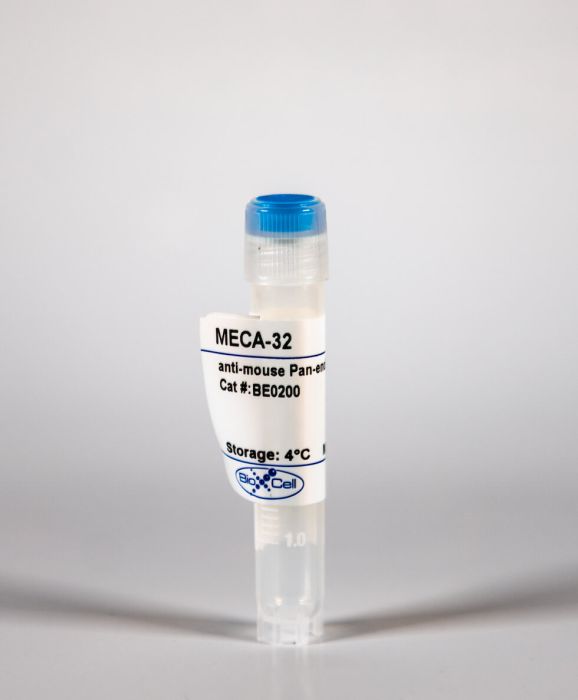
InVivoMAb anti-mouse Pan-endothelial Cell Antigen (MECA-32)
Product Details
The MECA-32 monoclonal antibody reacts with mouse pan-endothelial cell antigen also known as MECA-32 and PLVAP (plasmalemma vesicle-associated protein). MECA-32 is a 60 kDa transmembrane homodimer which is expressed on the surface of most endothelial cells. However, MECA-32 shows restricted distribution in the skeletal, cardiac and brain tissues of adult and embryonic mice. In embryonic mice, MECA-32 expression is developmentally regulated in the vessels associated with the blood brain barrier and persists only through day 16 of gestation.Specifications
| Isotype | Rat IgG2a, κ |
|---|---|
| Recommended Isotype Control(s) | InVivoMAb rat IgG2a isotype control, anti-trinitrophenol |
| Recommended Dilution Buffer | InVivoPure pH 7.0 Dilution Buffer |
| Immunogen | Mouse lymph node stromal cells |
| Reported Applications |
Immunohistochemistry (frozen) Flow cytometry Western blot Immunofluorescence |
| Formulation |
PBS, pH 7.0 Contains no stabilizers or preservatives |
| Endotoxin |
<2EU/mg (<0.002EU/μg) Determined by LAL gel clotting assay |
| Sterility | 0.2 μM filtered |
| Production | Purified from tissue culture supernatant in an animal free facility |
| Purification | Protein G |
| RRID | AB_10950167 |
| Molecular Weight | 150 kDa |
| Storage | The antibody solution should be stored at the stock concentration at 4°C. Do not freeze. |
Recommended Products
Flow Cytometry
Caveolae, fenestrae and transendothelial channels retain PV1 on the surface of endothelial cells PubMed
PV1 protein is an essential component of stomatal and fenestral diaphragms, which are formed at the plasma membrane of endothelial cells (ECs), on structures such as caveolae, fenestrae and transendothelial channels. Knockout of PV1 in mice results in in utero and perinatal mortality. To be able to interpret the complex PV1 knockout phenotype, it is critical to determine whether the formation of diaphragms is the only cellular role of PV1. We addressed this question by measuring the effect of complete and partial removal of structures capable of forming diaphragms on PV1 protein level. Removal of caveolae in mice by knocking out caveolin-1 or cavin-1 resulted in a dramatic reduction of PV1 protein level in lungs but not kidneys. The magnitude of PV1 reduction correlated with the abundance of structures capable of forming diaphragms in the microvasculature of these organs. The absence of caveolae in the lung ECs did not affect the transcription or translation of PV1, but it caused a sharp increase in PV1 protein internalization rate via a clathrin- and dynamin-independent pathway followed by degradation in lysosomes. Thus, PV1 is retained on the cell surface of ECs by structures capable of forming diaphragms, but undergoes rapid internalization and degradation in the absence of these structures, suggesting that formation of diaphragms is the only role of PV1.
Immunohistochemistry (frozen)
Paclitaxel therapy promotes breast cancer metastasis in a TLR4-dependent manner PubMed
Emerging evidence suggests that cytotoxic therapy may actually promote drug resistance and metastasis while inhibiting the growth of primary tumors. Work in preclinical models of breast cancer has shown that acquired chemoresistance to the widely used drug paclitaxel can be mediated by activation of the Toll-like receptor TLR4 in cancer cells. In this study, we determined the prometastatic effects of tumor-expressed TLR4 and paclitaxel therapy and investigated the mechanisms mediating these effects. While paclitaxel treatment was largely efficacious in inhibiting TLR4-negative tumors, it significantly increased the incidence and burden of pulmonary and lymphatic metastasis by TLR4-positive tumors. TLR4 activation by paclitaxel strongly increased the expression of inflammatory mediators, not only locally in the primary tumor microenvironment but also systemically in the blood, lymph nodes, spleen, bone marrow, and lungs. These proinflammatory changes promoted the outgrowth of Ly6C(+) and Ly6G(+) myeloid progenitor cells and their mobilization to tumors, where they increased blood vessel formation but not invasion of these vessels. In contrast, paclitaxel-mediated activation of TLR4-positive tumors induced de novo generation of deep intratumoral lymphatic vessels that were highly permissive to invasion by malignant cells. These results suggest that paclitaxel therapy of patients with TLR4-expressing tumors may activate systemic inflammatory circuits that promote angiogenesis, lymphangiogenesis, and metastasis, both at local sites and premetastatic niches where invasion occurs in distal organs. Taken together, our findings suggest that efforts to target TLR4 on tumor cells may simultaneously quell local and systemic inflammatory pathways that promote malignant progression, with implications for how to prevent tumor recurrence and the establishment of metastatic lesions, either during chemotherapy or after it is completed.
Immunofluorescence, Western Blot
Fetal liver endothelium regulates the seeding of tissue-resident macrophages PubMed
Macrophages are required for normal embryogenesis, tissue homeostasis and immunity against microorganisms and tumours. Adult tissue-resident macrophages largely originate from long-lived, self-renewing embryonic precursors and not from haematopoietic stem-cell activity in the bone marrow. Although fate-mapping studies have uncovered a great amount of detail on the origin and kinetics of fetal macrophage development in the yolk sac and liver, the molecules that govern the tissue-specific migration of these cells remain completely unknown. Here we show that an endothelium-specific molecule, plasmalemma vesicle-associated protein (PLVAP), regulates the seeding of fetal monocyte-derived macrophages to tissues in mice. We found that PLVAP-deficient mice have completely normal levels of both yolk-sac- and bone-marrow-derived macrophages, but that fetal liver monocyte-derived macrophage populations were practically missing from tissues. Adult PLVAP-deficient mice show major alterations in macrophage-dependent iron recycling and mammary branching morphogenesis. PLVAP forms diaphragms in the fenestrae of liver sinusoidal endothelium during embryogenesis, interacts with chemoattractants and adhesion molecules and regulates the egress of fetal liver monocytes to the systemic vasculature. Thus, PLVAP selectively controls the exit of macrophage precursors from the fetal liver and, to our knowledge, is the first molecule identified in any organ as regulating the migratory events during embryonic macrophage ontogeny.


