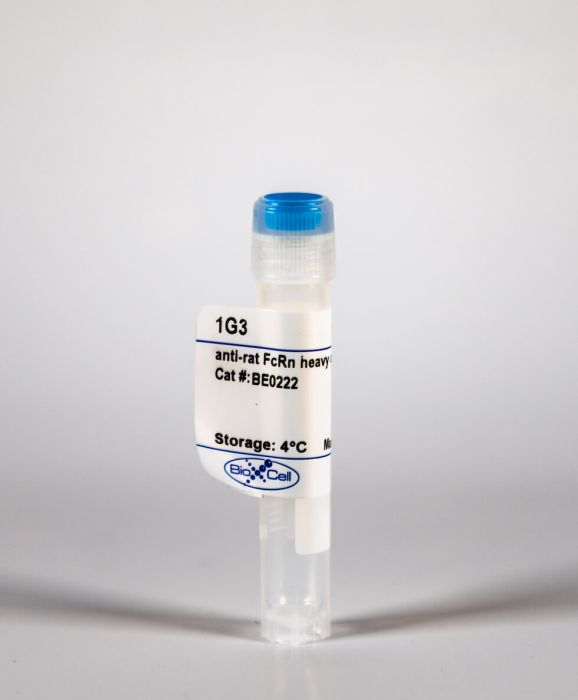
InVivoMAb anti-rat FcRn heavy chain heterodimers
Product Details
The 1G3 antibody was raised against soluble rat neonatal Fc receptor (FcRn) in an adjuvant. FcRn is a heterodimer composed of a membrane bound heavy chain attached non-covalently to β2-microgloublin. It is structurally similar to MHC class I molecules. The 1G3 antibody is used in studies of the MHC class I heavy chain FcRn heterodimers and their interaction with IgG.Specifications
| Isotype | Mouse IgG1 |
|---|---|
| Recommended Isotype Control(s) | InVivoMAb mouse IgG1 isotype control, unknown specificity |
| Recommended Dilution Buffer | InVivoPure pH 7.0 Dilution Buffer |
| Immunogen | Purified soluble FcRn |
| Reported Applications |
Immunofluorescence ELISA Flow cytometry |
| Formulation |
PBS, pH 7.0 Contains no stabilizers or preservatives |
| Endotoxin |
<2EU/mg (<0.002EU/μg) Determined by LAL gel clotting assay |
| Sterility | 0.2 μM filtered |
| Production | Purified from tissue culture supernatant in an animal free facility |
| Purification | Protein G |
| RRID | AB_2687705 |
| Molecular Weight | 150 kDa |
| Storage | The antibody solution should be stored at the stock concentration at 4°C. Do not freeze. |
Recommended Products
ELISA, Western Blot
Investigation of the interaction between the class I MHC-related Fc receptor and its immunoglobulin G ligand PubMed
The neonatal Fc receptor (FcRn) is structurally similar to class I major histocompatibility molecules. FcRn transports maternal immunoglobulin G (IgG) from ingested milk into the blood. IgG is bound at the pH of milk (pH 6.0-6.5) in the gut and released at the pH of blood (pH 7.5). We find that alteration of a histidine pair within the alpha 3 domain of FcRn and of a nearby loop (the FcRn counterpart of the class I CD8-binding loop) affects the affinity for IgG. Inhibition studies suggest the involvement of the FcRn B2-microglobulin domain in IgG binding. Fragment B of protein A inhibits FcRn binding to IgG, localizing the binding site on Fc for FcRn to the CH2-CH3 domain interface. Three histidines present at the CH2-CH3 domain interface of Fc could be partially responsible for the pH-dependent interaction between FcRn and IgG.
Immunofluorescence
Expression of the neonatal Fc receptor (FcRn) at the blood-brain barrier PubMed
The blood-brain barrier (BBB) restricts transport of immunoglobulin G (IgG) in the blood to brain direction. However, IgG undergoes rapid efflux in the brain to blood direction via reverse transcytosis across the BBB after direct intracerebral injection. This BBB IgG transport system has the characteristics of an Fc receptor (FcR), but there is no molecular information on the putative BBB FcR. The present study uses confocal microscopy and an antibody to the rat neonatal FcR (FcRn), and demonstrates the expression of the FcRn at the brain microvasculature and choroid plexus epithelium. Co-localization with the Glut1 glucose transporter indicates the brain microvascular FcRn is expressed in the capillary endothelium. The capillary endothelial FcRn may mediate the ‘reverse transcytosis’ of IgG in the brain to blood direction.
Immunofluorescence
Expression of neonatal Fc receptor in the eye PubMed
PURPOSE: The neonatal Fc receptor (FcRn) plays a critical role in the homeostasis and degradation of immunoglobulin G (IgG). It mediates the transport of IgG across epithelial cell barriers and recycles IgG in endothelial cells back into the bloodstream. These functions critically depend on the binding of FcRn to the Fc domain of IgG. The half-life and distribution of intravitreally injected anti-VEGF molecules containing IgG-Fc domains might therefore be affected by FcRn expressed in the eye. In order to establish whether FcRn-Fc(IgG) interactions may occur in the eye, we studied the mRNA and protein distribution of FcRn in postmortem ocular tissue. METHODS: We used qPCR to study mRNA expression of the transmembrane chain of FcRn (FCGRT) in retina, optic nerve, RPE/choroid plexus, ciliary body/iris plexus, lens, cornea, and conjunctiva isolated from mouse, rat, pig, and human postmortem eyes and used immunohistochemistry to determine the pattern of FcRn expression in FCGRT-transgenic mouse and human eyes. RESULTS: In all four tested species, Fcgrt mRNA was expressed in the retina, RPE/choroid, and the ciliary body/iris, while immunohistochemistry documented FcRn protein expression in the ciliary body epithelium, macrophages, and endothelial cells in the retinal and choroidal vasculature. CONCLUSIONS: Our results demonstrate that FcRn has the potential to interact with IgG-Fc domains in the ciliary epithelium and retinal and choroidal vasculature, which might affect the half-life and distribution of intravitreally injected Fc-carrying molecules.


