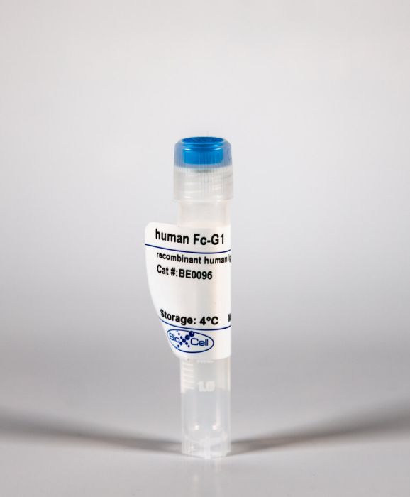
InVivoMAb recombinant human IgG1 Fc
Product Details
This recombinant human IgG1 Fc is the Fc fragment of human IgG1 only and does not contain the Fab fragments. The molecular mass of the recombinant human IgG1 Fc is approximately 34 kDa in SDS-PAGE under reducing conditions. This product is commonly used as an isotype control for human IgG1 antibodies as well as fusion proteins containing the human IgG Fc fragment.Specifications
| Recommended Dilution Buffer | InVivoPure pH 7.0 Dilution Buffer |
|---|---|
| Formulation |
PBS, pH 7.0 Contains no stabilizers or preservatives |
| Endotoxin |
<2EU/mg (<0.002EU/μg) Determined by LAL gel clotting assay |
| Sterility | 0.2 μM filtered |
| Production | Purified from tissue culture supernatant in an animal free facility |
| Purification | Protein A |
| RRID | AB_1107777 |
| Storage | The antibody solution should be stored at the stock concentration at 4°C. Do not freeze. |
Recommended Products
in vivo IFNγ neutralization
IFN-gamma dictates allograft fate via opposing effects on the graft and on recipient CD8 T cell responses PubMed
CD8 T cells are necessary for costimulation blockade-resistant rejection. However, the mechanism by which CD8 T cells mediate rejection in the absence of major costimulatory signals is poorly understood. IFN-gamma promotes CD8 T cell-mediated immune responses, but IFN-gamma-deficient mice show early graft loss despite costimulation blockade. In contrast, we found that IFN-gamma receptor knockout mice show dramatically prolonged graft survival under costimulation blockade. To investigate this paradox, we addressed the effects of IFN-gamma on T cell alloresponses in vivo independent of the effects of IFN-gamma on graft survival. We identified a donor-specific CD8 T cell breakthrough response temporally correlated with costimulation blockade-resistant rejection. Neither IFN-gamma receptor knockout recipients nor IFN-gamma-deficient recipients showed a CD8 breakthrough response. Graft death on IFN-gamma-deficient recipients despite costimulation blockade could be explained by the lack of IFN-gamma available to act on the graft. Indeed, the presence of IFN-gamma was necessary for graft survival on IFN-gamma receptor knockout recipients, as either IFN-gamma neutralization or the lack of the IFN-gamma receptor on the graft precipitated early graft loss. Thus, IFN-gamma is required both for the recipient to mount a donor-specific CD8 T cell response under costimulation blockade as well as for the graft to survive after allotransplantation.
Pharmacological Evaluation of the SCID T Cell Transfer Model of Colitis: As a Model of Crohn's Disease PubMed
Animal models are important tools in the development of new drug candidates against the inflammatory bowel diseases (IBDs) Crohn’s disease and ulcerative colitis. In order to increase the translational value of these models, it is important to increase knowledge relating to standard drugs. Using the SCID adoptive transfer colitis model, we have evaluated the effect of currently used IBD drugs and IBD drug candidates, that is, anti-TNF-alpha, TNFR-Fc, anti-IL-12p40, anti-IL-6, CTLA4-Ig, anti-alpha4beta7 integrin, enrofloxacin/metronidazole, and cyclosporine. We found that anti-TNF-alpha, antibiotics, anti-IL-12p40, anti-alpha4beta7 integrin, CTLA4-Ig, and anti-IL-6 effectively prevented onset of colitis, whereas TNFR-Fc and cyclosporine did not. In intervention studies, antibiotics, anti-IL-12p40, and CTLA4-Ig induced remission, whereas the other compounds did not. The data suggest that the adoptive transfer model and the inflammatory bowel diseases have some main inflammatory pathways in common. The finding that some well-established IBD therapeutics do not have any effect in the model highlights important differences between the experimental model and the human disease.
Immunofluorescence, in vitro IL-4 neutralization, in vitro T cell stimulation/activation
Regulation of autoimmune germinal center reactions in lupus-prone BXD2 mice by follicular helper T cells PubMed
BXD2 mice spontaneously develop autoantibodies and subsequent glomerulonephritis, offering a useful animal model to study autoimmune lupus. Although initial studies showed a critical contribution of IL-17 and Th17 cells in mediating autoimmune B cell responses in BXD2 mice, the role of follicular helper T (Tfh) cells remains incompletely understood. We found that both the frequency of Th17 cells and the levels of IL-17 in circulation in BXD2 mice were comparable to those of wild-type. By contrast, the frequency of PD-1+ CXCR5+ Tfh cells was significantly increased in BXD2 mice compared with wild-type mice, while the frequency of PD-1+ CXCR5+ Foxp3+ follicular regulatory T (Tfr) cells was reduced in the former group. The frequency of Tfh cells rather than that of Th17 cells was positively correlated with the frequency of germinal center B cells as well as the levels of autoantibodies to dsDNA. More importantly, CXCR5+ CD4+ T cells isolated from BXD2 mice induced the production of IgG from naive B cells in an IL-21-dependent manner, while CCR6+ CD4+ T cells failed to do so. These results together demonstrate that Tfh cells rather than Th17 cells contribute to the autoimmune germinal center reactions in BXD2 mice.
in vivo blocking of IL-10/IL-10R signaling, in vivo CD4+ T cell depletion, in vivo CD8+ T cell depletion, in vivo regulatory T cell depletion, in vivo TNFα neutralization
Depletion of regulatory T cells in a hapten-induced inflammation model results in prolonged and increased inflammation driven by T cells PubMed
Regulatory T cells (Tregs ) are known to play an immunosuppressive role in the response of contact hypersensitivity (CHS), but neither the dynamics of Tregs during the CHS response nor the exaggerated inflammatory response after depletion of Tregs has been characterized in detail. In this study we show that the number of Tregs in the challenged tissue peak at the same time as the ear-swelling reaches its maximum on day 1 after challenge, whereas the number of Tregs in the draining lymph nodes peaks at day 2. As expected, depletion of Tregs by injection of a monoclonal antibody to CD25 prior to sensitization led to a prolonged and sustained inflammatory response which was dependent upon CD8 T cells, and co-stimulatory blockade with cytotoxic T lymphocyte antigen-4-immunoglobulin (CTLA-4-Ig) suppressed the exaggerated inflammation. In contrast, blockade of the interleukin (IL)-10-receptor (IL-10R) did not further increase the exaggerated inflammatory response in the Treg -depleted mice. In the absence of Tregs , the response changed from a mainly acute reaction with heavy infiltration of neutrophils to a sustained response with more chronic characteristics (fewer neutrophils and dominated by macrophages). Furthermore, depletion of Tregs enhanced the release of cytokines and chemokines locally in the inflamed ear and augmented serum levels of the systemic inflammatory mediators serum amyloid (SAP) and haptoglobin early in the response.
Niche-specific MHC II and PD-L1 regulate CD4+CD8αα+ intraepithelial lymphocyte differentiation PubMed
Conventional CD4+ T cells are differentiated into CD4+CD8αα+ intraepithelial lymphocytes (IELs) in the intestine; however, the roles of intestinal epithelial cells (IECs) are poorly understood. Here, we showed that IECs expressed MHC class II (MHC II) and programmed death-ligand 1 (PD-L1) induced by the microbiota and IFN-γ in the distal part of the small intestine, where CD4+ T cells were transformed into CD4+CD8αα+ IELs. Therefore, IEC-specific deletion of MHC II and PD-L1 hindered the development of CD4+CD8αα+ IELs. Intracellularly, PD-1 signals supported the acquisition of CD8αα by down-regulating the CD4-lineage transcription factor, T helper-inducing POZ/Krüppel-like factor (ThPOK), via the Src homology 2 domain-containing tyrosine phosphatase (SHP) pathway. Our results demonstrate that noncanonical antigen presentation with cosignals from IECs constitutes niche adaptation signals to develop tissue-resident CD4+CD8αα+ IELs.


