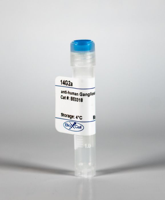
InVivoMAb anti-human Ganglioside GD2
Product Details
The 14G2a monoclonal antibody reacts with human ganglioside GD2 a sialic-acid bearing glycolipid that is involved in mediating cell attachment to the extracellular matrix. Ganglioside GD2 is expressed on tumors of neuroectodermal origin including human neuroblastoma and melanoma. The tumor specific expression of GD2 makes it a suitable target for immunotherapy with monoclonal antibodies or with artificial T cell receptors. Clone 14G2a is an isotype switch variant selected from the parental IgG3-producing hybridoma 14.18 and has identical reactivity as the parental antibody.Specifications
| Isotype | Mouse IgG2a, κ |
|---|---|
| Recommended Isotype Control(s) | InVivoMAb mouse IgG2a isotype control, unknown specificity |
| Recommended Dilution Buffer | InVivoPure pH 7.0 Dilution Buffer |
| Immunogen | Neuroblastoma cell line LAN-1 |
| Reported Applications |
in vitro induction of apoptosis in GD2+ cells in vivo inhibition of GD2+ tumor cell growth |
| Formulation |
PBS, pH 7.0 Contains no stabilizers or preservatives |
| Endotoxin |
<2EU/mg (<0.002EU/μg) Determined by LAL gel clotting assay |
| Sterility | 0.2 μM filtered |
| Production | Purified from tissue culture supernatant in an animal free facility |
| Purification | Protein G |
| RRID | AB_2819045 |
| Molecular Weight | 150 kDa |
| Storage | The antibody solution should be stored at the stock concentration at 4°C. Do not freeze. |
Recommended Products
in vivo induction of apoptosis in GD2+ cells
Functional properties and effect on growth suppression of human neuroblastoma tumors by isotype switch variants of monoclonal antiganglioside GD2 antibody 14.18 PubMed
A complete family of IgG isotype switch variant hybridomas was generated from the anti-GD2 monoclonal IgG3-producing hybridoma, 14.18, with the aid of the fluorescence-activated cell sorter. The IgG1, IgG2b, and IgG2a monoclonal antibodies (Mabs) produced by respective isotype switch variant hybridomas 14G1, 14G2b, or 14G2a, have binding activities for the biochemically defined GD2 antigen and GD2-expressing neuroblastoma target cell lines identical to that of IgG3 Mabs produced by the 14.18 parent cell line. This permitted us to examine the relative in vitro and in vivo cytotoxic capacities of each of the anti-GD2 antibodies for GD2-expressing neuroblastoma cells independent of antibody binding affinity or specificity. Mabs produced by 14.18, 14G2a, or 14G2b, but not 14G1, can direct efficient complement-dependent cytotoxicity against neuroblastoma tumor cells in the presence of human complement. Mabs produced by the parent 14.18 or by 14G2a are more efficient in directing antibody-dependent cell-mediated cytotoxicity than Mabs produced by 14G2b, and Mabs of 14G1 are inactive. However, despite these noted in vitro differences, antibodies produced by each member of this switch variant family suppress the growth of human neuroblastoma tumor cells in BALB/c athymic nu/nu mice. These studies suggest that a mechanism(s) other than Fc-directed complement-dependent cytotoxicity or antibody-dependent cell-mediated cytotoxicity may account for the in vivo antitumor effects of these particular antibodies.
in vitro induction of apoptosis in GD2+ cells
The GD2-specific 14G2a monoclonal antibody induces apoptosis and enhances cytotoxicity of chemotherapeutic drugs in IMR-32 human neuroblastoma cells PubMed
Neuroblastoma (NB) is the most common extracranial solid tumor of childhood. The majority of children suffers from high risk neuroblastoma and has disseminated disease at the time of diagnosis. Despite recent advances in chemotherapy, the prognoses for children with high risk NB remain poor. Therefore, new treatment modalities are urgently needed. GD2 ganglioside is an antigen that is highly expressed on NB cells with only limited distribution on healthy tissues. Consequently, it appears to be an ideal target for both active and passive immunotherapy. The immunological effector mechanisms mediated by anti-GD2 monoclonal antibodies (mAbs) have been already well characterized. However, a growing number of reports suggest that GD2-specific antibodies may exhibit anti-proliferative effects without the immune system involvement. Here, we have shown that anti-GD2 14G2a mAb is capable of decreasing survival of IMR-32 human neuroblastoma cells in a dose-dependent manner. Death induced by this antibody exhibited several characteristics typical for apoptosis such as increased number of Annexin V- and propidium iodide-positive cells, cleavage of caspase 3 and prominent rise in caspase activity. The use of a pan caspase inhibitor Z-VAD-fmk suggested that the killing potential of this mAb is partially caspase-dependent. 14G2a mAb was rapidly endocytosed upon antigen binding. Employment of chloroquine, an inhibitor of lysosomal degradation, did not rescue IMR-32 cells from antibody-induced cell death suggesting lack of ceramide involvement in the observed effect. Most importantly, our studies showed that at particular drug concentrations 14G2a mAb exerts a synergistic effect with doxorubicin and topotecan, as well as an additive effect with carboplatin in killing IMR-32 cells in vitro. Our results provide guidance regarding how to best combine GD2-specific 14G2a antibody with existing cancer therapeutic agents to improve available treatment modalities for neuroblastoma.
in vitro induction of apoptosis in GD2+ cells
GD2 ganglioside-binding antibody 14G2a and specific aurora A kinase inhibitor MK-5108 induce autophagy in IMR-32 neuroblastoma cells PubMed
The process of autophagy and its role in survival of human neuroblastoma cell cultures was studied upon addition of an anti-GD2 ganglioside (GD2) 14G2a mouse monoclonal antibody (14G2a mAb) and an aurora A kinase specific inhibitor, MK-5108. It was recently shown that combination of these agents significantly potentiates cytotoxicity against IMR-32 and CHP-134 neuroblastoma cells in vitro, as compared to the inhibitor used alone. In this study we gained mechanistic insights on autophagy in the observed cytotoxic effects exerted by both agents using cytotoxicity assays, RT-qPCR, immunoblotting, and autophagy detection methods. Enhancement of the autophagy process in the 14G2a mAb- and MK-5108-treated IMR-32 cells was documented by assessing autophagic flux. Application of a lysosomotropic agent-chloroquine (CQ) affected the 14G2a mAb- and MK-5108-stimulated autophagic flux. It is our conclusion that the 14G2a mAb (40 mug/ml) and MK-5108 inhibitor (0.1 muM) induce autophagy in IMR-32 cells. Moreover, the combinatorial treatment of IMR-32 cells with the 14G2a mAb and CQ significantly potentiates cytotoxic effect, as compared to CQ used alone. Most importantly, we showed that interfering with autophagy at its early and late step augments the 14G2a mAb-induced apoptosis, therefore we can conclude that inhibition of autophagy is the primary mechanism of the CQ-mediated sensitization to the 14G2a mAb-induced apoptosis. Although, there was no virtual stimulation of autophagy in the 14G2a mAb-treated CHP-134 neuroblastoma cells, we were able to show that PHLDA1 protein positively regulates autophagy and this process exists in a mutually exclusive manner with apoptosis in PHLDA1-silenced CHP-134 cells.


