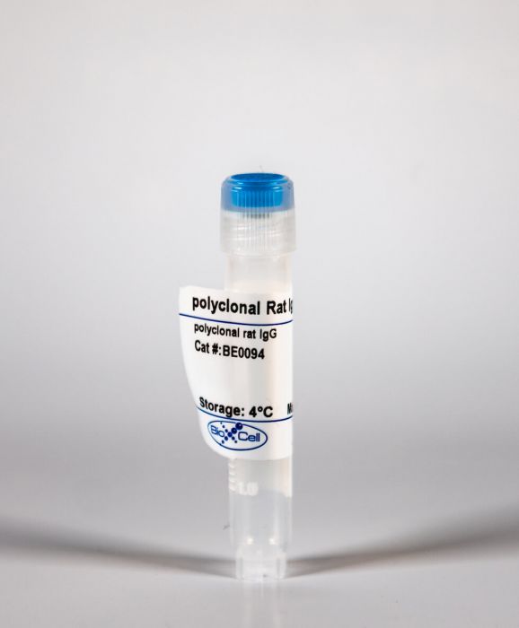
InVivoMAb polyclonal rat IgG
Product Details
The polyclonal rat IgG is purified from rat serum. It is ideal for use as a non-reactive control IgG for polyclonal rat IgG antibodies in most in vivo and in vitro applications.Specifications
| Isotype | Rat IgG |
|---|---|
| Recommended Dilution Buffer | InVivoPure pH 7.0 Dilution Buffer |
| Formulation |
PBS, pH 7.0 Contains no stabilizers or preservatives |
| Endotoxin |
<2EU/mg (<0.002EU/μg) Determined by LAL gel clotting assay |
| Sterility | 0.2 µm filtration |
| Production | Purification from rat serum |
| Purification | Protein G |
| RRID | AB_1107795 |
| Molecular Weight | 150 kDa |
| Storage | The antibody solution should be stored at the stock concentration at 4°C. Do not freeze. |
Recommended Products
in vitro IL-2 neutralization
In vitro-differentiated TH17 cells mediate lethal acute graft-versus-host disease with severe cutaneous and pulmonary pathologic manifestations PubMed
The morbidity and mortality associated with graft-host-disease (GVHD) is a significant obstacle to the greater use of allogeneic stem cell transplantation. Donor T cells that predominantly differentiate into TH1/Tc1 T cells and generate pro-inflammatory cytokines such as interferon-gamma (IFN-gamma) mediate GVHD. Although numerous studies have described a pathogenic role for IFN-gamma, multiple reports have demonstrated that the lack of IFN-gamma paradoxically exacerbated GVHD lethality. This has led to speculation that another subset of T cells may significantly contribute to GVHD mortality. Several groups have demonstrated a new lineage of CD4+ T helper cell development distinct from TH1 or TH2 differentiation. This lineage is characterized by production of interleukin (IL)-17A, IL-17F, IL-22, and IL-21 and has been termed TH17 cells. Here, we demonstrate that a highly purified population of TH17 cells is capable of inducing lethal GVHD, hallmarked by extensive pathologic cutaneous and pulmonary lesions. Upon transfer, these cells migrate to and expand in GVHD target organs and secondary lymphoid tissues. Finally, we demonstrate differential roles for tumor necrosis factor-alpha (TNF-alpha) and IL-17A in the clinical manifestations of GVHD induced by TH17 cells. Our studies demonstrate that cells other than TH1/Tc1 can mediate acute GVHD.
IL-33 expands suppressive CD11b+ Gr-1(int) and regulatory T cells, including ST2L+ Foxp3+ cells, and mediates regulatory T cell-dependent promotion of cardiac allograft survival PubMed
IL-33 administration is associated with facilitation of Th2 responses and cardioprotective properties in rodent models. However, in heart transplantation, the mechanism by which IL-33, signaling through ST2L (the membrane-bound form of ST2), promotes transplant survival is unclear. We report that IL-33 administration, while facilitating Th2 responses, also increases immunoregulatory myeloid cells and CD4(+) Foxp3(+) regulatory T cells (Tregs) in mice. IL-33 expands functional myeloid-derived suppressor cells, CD11b(+) cells that exhibit intermediate (int) levels of Gr-1 and potent T cell suppressive function. Furthermore, IL-33 administration causes an St2-dependent expansion of suppressive CD4(+) Foxp3(+) Tregs, including an ST2L(+) population. IL-33 monotherapy after fully allogeneic mouse heart transplantation resulted in significant graft prolongation associated with increased Th2-type responses and decreased systemic CD8(+) IFN-gamma(+) cells. Also, despite reducing overall CD3(+) cell infiltration of the graft, IL-33 administration markedly increased intragraft Foxp3(+) cells. Whereas control graft recipients displayed increases in systemic CD11b(+) Gr-1(hi) cells, IL-33-treated recipients exhibited increased CD11b(+) Gr-1(int) cells. Enhanced ST2 expression was observed in the myocardium and endothelium of rejecting allografts, however the therapeutic effect of IL-33 required recipient St2 expression and was dependent on Tregs. These findings reveal a new immunoregulatory property of IL-33. Specifically, in addition to supporting Th2 responses, IL-33 facilitates regulatory cells, particularly functional CD4(+) Foxp3(+) Tregs that underlie IL-33-mediated cardiac allograft survival.
PD-L1-expressing dendritic cells contribute to viral resistance during acute HSV-1 infection PubMed
The inhibitory receptor, Programmed Death 1 (PD-1), and its ligands (PD-L1/PD-L2) are thought to play a role in immune surveillance during chronic viral infection. The contribution of the receptor/ligand pair during an acute infection is less understood. To determine the role of PD-L1 and PD-L2 during acute ocular herpes simplex virus type 1 (HSV-1) infection, HSV-1-infected mice administered neutralizing antibody to PD-L1 or PD-L2 were assessed for viral burden and host cellular immune responses. Virus titers were elevated in cornea and trigeminal ganglia (TG) of anti-PD-L1-treated mice which corresponded with a reduced number of CD80-expressing dendritic cells, PD-L1(+) dendritic cells, and HSV-1-specific CD8(+) T cells within the draining (mandibular) lymph node (MLN). In contrast, anti-PD-L2 treatment had no effect on viral replication or changes in the MLN population. Notably, analysis of CD11c-enriched MLN cells from anti-PD-L1-treated mice revealed impaired functional capabilities. These studies indicate PD-L1-expressing dendritic cells are important for antiviral defense during acute HSV-1 infection.
CXCR1 and CXCR2 Chemokine Receptor Agonists Produced by Tumors Induce Neutrophil Extracellular Traps that Interfere with Immune Cytotoxicity PubMed
Neutrophils are expanded and abundant in cancer-bearing hosts. Under the influence of CXCR1 and CXCR2 chemokine receptor agonists and other chemotactic factors produced by tumors, neutrophils, and granulocytic myeloid-derived suppressor cells (MDSCs) from cancer patients extrude their neutrophil extracellular traps (NETs). In our hands, CXCR1 and CXCR2 agonists proved to be the major mediators of cancer-promoted NETosis. NETs wrap and coat tumor cells and shield them from cytotoxicity, as mediated by CD8(+) T cells and natural killer (NK) cells, by obstructing contact between immune cells and the surrounding target cells. Tumor cells protected from cytotoxicity by NETs underlie successful cancer metastases in mice and the immunotherapeutic synergy of protein arginine deiminase 4 (PAD4) inhibitors, which curtail NETosis with immune checkpoint inhibitors. Intravital microscopy provides evidence of neutrophil NETs interfering cytolytic cytotoxic T lymphocytes (CTLs) and NK cell contacts with tumor cells.
Decreased NSG3 enhances PD-L1 expression by Erk1/2 pathway to promote pancreatic cancer progress PubMed
Inhibiting the functioning of PD-1/PD-L1 to activate human immune system and improve the prognosis of pancreatic cancer (PC) would provide a significant boost to handling the disease. One research found the expression level of NSG3 was reduced in pediatric pilocytic astrocytoma, so is PC and we found NSG3 could regulate the expression of PD-L1. So NSG3 could become a new target for enhancing the immune response to PC. The GEPIA website was employed to analyze the prognoses in PC patients with different NSG3 levels. Immunohistochemistry (IHC) analysis was applied to detect different levels of NSG3 in para-PC and PC tissues. Cell biological function tests (in vitro) were performed and a subcutaneous nude mice tumor model (in vivo) was established to verify the effect of NSG3 on PC. Immunoblotting and RT-qPCR were utilized to demonstrate the inhibiting effect of NSG3 on PD-L1 through regulating Erk1/2 phosphorylation. A subcutaneous C57BL/6 tumor mice model was established to assess the possibility of a synergistic effect of NSG3 expression and the use of an anti-PD-L1 antibody on PC. PC tissues had decreased NSG3 expression levels, which led to poor prognosis. Overexpressing NSG3 suppressed proliferation, invasion and migration capacities of PC cells. On the contrary, knocking-down NSG3 prompted PC malignancy whether in vivo or in vitro. Importantly, NSG3 prevented Erk1/2 phosphorylation to inhibit PD-L1 expression. Additionally, NSG3 and an immune checkpoint inhibitor anti-PD-1 antibody acted synergistically, which enhanced the efficacy of the inhibitor. NSG3 inhibited PD-L1 expression by suppressing Erk1/2 phosphorylation to improve the immune response to PC. NSG3 is, therefore, a potential new diagnostic and prognostic marker, particularly useful in immune checkpoint blockade therapy.


