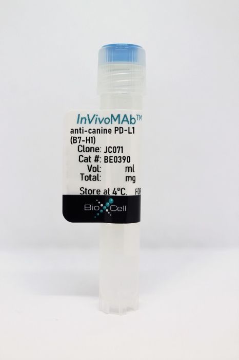
InVivoMAb anti-canine PD-L1 (B7-H1)
Product Details
The JC071 monoclonal antibody reacts with canine PD-L1 (programmed death ligand 1) also known as B7-H1 or CD274. PD-L1 is a 40 kDa type I transmembrane protein that belongs to the B7 family of the Ig superfamily. PD-L1 is expressed on T lymphocytes, B lymphocytes, NK cells, dendritic cells, as well as IFNγ stimulated monocytes, epithelial cells, and endothelial cells. PD-L1 binds to its receptor, PD-1, found on CD4 and CD8 thymocytes as well as activated T and B lymphocytes and myeloid cells. Engagement of PD-L1 with PD-1 leads to inhibition of TCR-mediated T cell proliferation and cytokine production. PD-L1 is thought to play an important role in tumor immune evasion. Induced PD-L1 expression is common in many tumors and results in increased resistance of tumor cells to CD8 T cell mediated lysis. The JC071 antibody can detect PD-L1 on canine tissues by flow cytometry and Western blot.Specifications
| Isotype | Mouse IgG1, κ |
|---|---|
| Recommended Isotype Control(s) | InVivoMAb mouse IgG1 isotype control, unknown specificity |
| Recommended Dilution Buffer | InVivoPure pH 7.0 Dilution Buffer |
| Immunogen | Recombinant canine PD-L1-Ig fusion protein |
| Reported Applications |
in vitro PD-L1 blockade Functional assays Western blot Flow cytometry |
| Formulation |
PBS, pH 7.0 Contains no stabilizers or preservatives |
| Endotoxin |
<2EU/mg (<0.002EU/μg) Determined by LAL gel clotting assay |
| Sterility | 0.2 µm filtration |
| Production | Purified from tissue culture supernatant in an animal free facility |
| Purification | Protein G |
| Molecular Weight | 150 kDa |
| Storage | The antibody solution should be stored at the stock concentration at 4°C. Do not freeze. |
Recommended Products
Flow Cytometry, Functional assays, in vitro PD-L1 blockade, Western Blot
Development of canine PD-1/PD-L1 specific monoclonal antibodies and amplification of canine T cell function PubMed
Interruption of the programmed death 1 (PD-1) / programmed death ligand 1 (PD-L1) pathway is an established and effective therapeutic strategy in human oncology and holds promise for veterinary oncology. We report the generation and characterization of monoclonal antibodies specific for canine PD-1 and PD-L1. Antibodies were initially assessed for their capacity to block the binding of recombinant canine PD-1 to recombinant canine PD-L1 and then ranked based on efficiency of binding as judged by flow cytometry. Selected antibodies were capable of detecting PD-1 and PD-L1 on canine tissues by flow cytometry and Western blot. Anti-PD-L1 worked for immunocytochemistry and anti-PD-1 worked for immunohistochemistry on formalin-fixed paraffin embedded canine tissues, suggesting the usage of this antibody with archived tissues. Additionally, anti-PD-L1 (JC071) revealed significantly increased PD-L1 expression on canine monocytes after stimulation with peptidoglycan or lipopolysaccharide. Together, these antibodies display specificity for the natural canine ligand using a variety of potential diagnostic applications. Importantly, multiple PD-L1-specific antibodies amplified IFN-γ production in a canine peripheral blood mononuclear cells (PBMC) concanavlin A (Con A) stimulation assay, demonstrating functional activity.


