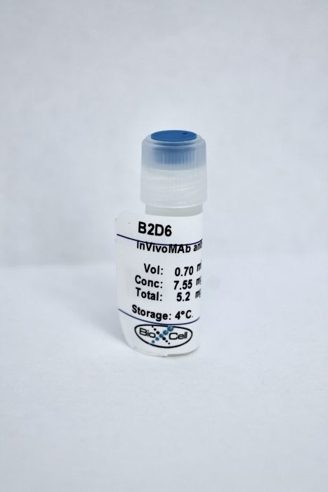
InVivoMAb anti-human EphA2
Product Details
The B2D6 monoclonal antibody reacts with human Ephrin type-A receptor 2 (EphA2). EphA2 is a ~130 kDa type I transmembrane glycoprotein that belongs to the receptor tyrosine kinase family. EphA2 is variably expressed by epithelial cells, dendritic cells, Langerhans cells, keratinocytes, and endothelial cells. EphA2 functions as a receptor for glycophosphatidylinositol (GPI) membrane-linked members of the Ephrin-A family, including Ephrins A1-A5. EphA2 is involved in regulating cellular growth, adhesion, migration, survival, and plays a role in angiogenesis. Its expression may be upregulated on vascular endothelium in certain breast, prostate, and colon cancers as well as on some metastatic tumor cells.Specifications
| Isotype | Mouse IgG2b, κ |
|---|---|
| Recommended Isotype Control(s) | InVivoMAb mouse IgG2b isotype control, unknown specificity |
| Recommended Dilution Buffer | InVivoPure pH 7.0 Dilution Buffer |
| Immunogen | Human EphA2 isolated from Ras-transformed MCF-10A cells |
| Reported Applications |
Immunohistochemistry (paraffin) Immunoprecipitation Functional assay |
| Formulation |
PBS, pH 7.0 Contains no stabilizers or preservatives |
| Endotoxin |
<2EU/mg (<0.002EU/μg) Determined by LAL gel clotting assay |
| Sterility | 0.2 μM filtered |
| Production | Purified from tissue culture supernatant in an animal free facility |
| Purification | Protein A |
| RRID | AB_2894761 |
| Molecular Weight | 150 kDa |
| Storage | The antibody solution should be stored at the stock concentration at 4°C. Do not freeze. |
Recommended Products
Functional assays, Immunoprecipitation
E-cadherin regulates the function of the EphA2 receptor tyrosine kinase PubMed
EphA2 is a member of the Eph family of receptor tyrosine kinases, which are increasingly understood to play critical roles in disease and development. We report here the regulation of EphA2 by E-cadherin. In nonneoplastic epithelia, EphA2 was tyrosine-phosphorylated and localized to sites of cell-cell contact. These properties required the proper expression and functioning of E-cadherin. In breast cancer cells that lack E-cadherin, the phosphotyrosine content of EphA2 was decreased, and EphA2 was redistributed into membrane ruffles. Expression of E-cadherin in metastatic cells restored a more normal pattern of EphA2 phosphorylation and localization. Activation of EphA2, either by E-cadherin expression or antibody-mediated aggregation, decreased cell-extracellular matrix adhesion and cell growth. Altogether, this demonstrates that EphA2 function is dependent on E-cadherin and suggests that loss of E-cadherin function may alter neoplastic cell growth and adhesion via effects on EphA2.
Immunohistochemistry (paraffin)
EphA2 overexpression causes tumorigenesis of mammary epithelial cells PubMed
Elevated levels of protein tyrosine phosphorylation contribute to a malignant phenotype, although the tyrosine kinases that are responsible for this signaling remain largely unknown. Here we report increased levels of the EphA2 (ECK) protein tyrosine kinase in clinical specimens and cell models of breast cancer. We also show that EphA2 overexpression is sufficient to confer malignant transformation and tumorigenic potential on nontransformed (MCF-10A) mammary epithelial cells. The transforming capacity of EphA2 is related to the failure of EphA2 to interact with its cell-attached ligands. Interestingly, stimulation of EphA2 reverses the malignant growth and invasiveness of EphA2-transformed cells. Taken together, these results identify EphA2 as a powerful oncoprotein in breast cancer.
Immunohistochemistry (paraffin)
VE-cadherin regulates EphA2 in aggressive melanoma cells through a novel signaling pathway: implications for vasculogenic mimicry PubMed
The formation of matrix-rich, vasculogenic-like networks, termed vasculogenic mimicry (VM), is a unique process characteristic of highly aggressive melanoma cells found to express genes previously thought to be exclusively associated with endothelial and epithelial cells. This study contributes new observations demonstrating that VE-cadherin can regulate the expression of EphA2 at the cell membrane by mediating its ability to become phosphorylated through interactions with its membrane bound ligand, ephrin-A1. VE-cadherin and EphA2 were also found to be colocalized in cell-cell adhesion junctions, both in vitro and in vivo. Immunoprecipitation studies revealed that EphA2 and VE-cadherin could interact, directly and/or indirectly, during VM. Furthermore, there was no change in the colocalization of EphA2 and VE-cadherin at cell-cell adhesion sites when EphA2 was phosphorylated on tyrosine residues. Although transient knockout of EphA2 expression did not alter VE-cadherin localization, transient knockout of VE-cadherin expression resulted in the reorganization of EphA2 on the cells’ surface, an accumulation of EphA2 in the cytoplasm, and subsequent dephosphorylation of EphA2. Collectively, these results suggest that VE-cadherin and EphA2 act in a coordinated manner as a key regulatory element in the process of melanoma VM and illuminate a novel signaling pathway that could be potentially exploited for therapeutic intervention.


