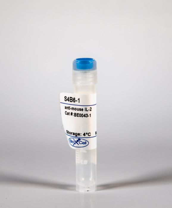
InVivoMAb anti-mouse IL-2
Product Details
The S4B6-1 monoclonal antibody reacts with mouse IL-2, a 17 kDa cytokine that is mainly produced by T cells in response to antigenic or mitogenic stimulation. IL-2 is required for T cell proliferation and other activities crucial to the regulation of immunity. The cytokine can also stimulate the growth and differentiation of B cells, monocytes/macrophages, and NK cells. Additionally, IL-2 prevents autoimmune diseases by promoting the differentiation of certain immature T cells into regulatory T cells. The S4B6-1 antibody has been shown to neutralize IL-2 in vivo.Specifications
| Isotype | Rat IgG2a |
|---|---|
| Recommended Isotype Control(s) | InVivoMAb rat IgG2a isotype control, anti-trinitrophenol |
| Recommended Dilution Buffer | InVivoPure pH 8.0 Dilution Buffer |
| Immunogen | Recombinant mouse IL-2 |
| Reported Applications |
in vivo IL-2 neutralization in vivo IL-2 receptor stimulation (as a complex with IL-2) |
| Formulation |
PBS, pH 8.0 Contains no stabilizers or preservatives |
| Endotoxin |
<2EU/mg (<0.002EU/μg) Determined by LAL gel clotting assay |
| Sterility | 0.2 μM filtered |
| Production | Purified from tissue culture supernatant in an animal free facility |
| Purification | Protein G |
| Molecular Weight | 150 kDa |
| Storage | The antibody solution should be stored at the stock concentration at 4°C. Do not freeze. |
Recommended Products
in vivo IL-2 receptor stimulation (as a complex with IL-2)
IL-2 complex treatment can protect naive mice from bacterial and viral infection PubMed
IL-2 complexes have substantial effects on the cellular immune system, and this approach is being explored for therapeutic application in infection and cancer. However, the impact of such treatments on subsequent encounter with pathogens has not been investigated. In this study, we report that naive mice treated with a short course of IL-2 complexes show enhanced protection from newly encountered bacterial and viral infections. IL-2 complex treatment expands both the NK and CD8 memory cell pool, including a recently described population of preexisting memory-phenotype T cells responsive to previously unencountered foreign Ags. Surprisingly, prolonged IL-2 complex treatment decreased CD8 T cell function and protective immunity. These data reveal the impact of cytokine complex treatment on the primary response to infection.
in vivo IL-2 receptor stimulation (as a complex with IL-2)
Differential outcome of IL-2/anti-IL-2 complex therapy on effector and memory CD8+ T cells following vaccination with an adenoviral vector encoding EBV epitopes PubMed
IL-2/anti-IL-2 complex-based therapy has been proposed as a potential adjunct therapeutic tool to enhance in vivo efficacy of T cell-based immunotherapeutic strategies for chronic viral infections and human cancers. In this study, we demonstrate that IL-2 complex therapy can have discerning effects on CD8(+) T cells depending on their stage of differentiation. To delineate the underlying mechanism for these opposing effects on CD8(+) T cells, we examined the effects of IL-2 therapy during early priming, effector, and memory phases of T cell responses generated following immunization with an adenoviral vector encoding multiple EBV CD8(+) epitopes. IL-2 complex treatment during the early priming phase, which coincided with low levels of IL-2Rbeta (CD122) and higher levels of IL-2Ralpha (CD25) on CD8(+) T cells, did not induce the expansion of effector T cells. In contrast, IL-2 complex treatment following the establishment of memory enhanced the expansion of Ag-specific T cells. Additionally, central memory T cells preferentially expanded following treatment at the expense of effector memory T cell populations. These studies demonstrate how differentiation status of the responding CD8(+) T cells impacts on their responsiveness to IL-2 complexes and highlight that timing of treatment should be considered before implementing this therapy in a clinical setting.
in vivo IL-2 neutralization, in vivo IL-2 receptor stimulation (as a complex with IL-2)
IL-2-dependent adaptive control of NK cell homeostasis PubMed
Activation and expansion of T and B lymphocytes and myeloid cells are controlled by Foxp3(+) regulatory T cells (T reg cells), and their deficiency results in a fatal lympho- and myeloproliferative syndrome. A role for T reg cells in the homeostasis of innate lymphocyte lineages remained unknown. Here, we report that T reg cells restrained the expansion of immature CD127(+) NK cells, which had the unique ability to up-regulate the IL2Ralpha (CD25) in response to the proinflammatory cytokine IL-12. In addition, we observed the preferential accumulation of CD127(+) NK cells in mice bearing progressing tumors or suffering from chronic viral infection. CD127(+) NK cells expanded in an IL-2-dependent manner upon T reg cell depletion and were able to give rise to mature NK cells, indicating that the latter can develop through a CD25(+) intermediate stage. Thus, T reg cells restrain the IL-2-dependent CD4(+) T cell help for CD127(+) immature NK cells. These findings highlight the adaptive control of innate lymphocyte homeostasis.
in vivo blocking of IL-7Rα signaling, in vivo IL-2 neutralization
Cutting Edge: memory regulatory t cells require IL-7 and not IL-2 for their maintenance in peripheral tissues PubMed
Thymic Foxp3-expressing regulatory T cells are activated by peripheral self-antigen to increase their suppressive function, and a fraction of these cells survive as memory regulatory T cells (mTregs). mTregs persist in nonlymphoid tissue after cessation of Ag expression and have enhanced capacity to suppress tissue-specific autoimmunity. In this study, we show that murine mTregs express specific effector memory T cell markers and localize preferentially to hair follicles in skin. Memory Tregs express high levels of both IL-2Ralpha and IL-7Ralpha. Using a genetic-deletion approach, we show that IL-2 is required to generate mTregs from naive CD4(+) T cell precursors in vivo. However, IL-2 is not required to maintain these cells in the skin and skin-draining lymph nodes. Conversely, IL-7 is essential for maintaining mTregs in skin in the steady state. These results elucidate the fundamental biology of mTregs and show that IL-7 plays an important role in their survival in skin.
in vivo CD80 blockade, In vivo CD86 blockade, in vivo ICOSL neutralization, in vivo IL-2 receptor stimulation (as a complex with IL-2)
Type I interferons directly inhibit regulatory T cells to allow optimal antiviral T cell responses during acute LCMV infection PubMed
Regulatory T (T reg) cells play an essential role in preventing autoimmunity but can also impair clearance of foreign pathogens. Paradoxically, signals known to promote T reg cell function are abundant during infection and could inappropriately enhance T reg cell activity. How T reg cell function is restrained during infection to allow the generation of effective antiviral responses remains largely unclear. We demonstrate that the potent antiviral type I interferons (IFNs) directly inhibit co-stimulation-dependent T reg cell activation and proliferation, both in vitro and in vivo during acute infection with lymphocytic choriomeningitis virus (LCMV). Loss of the type I IFN receptor specifically in T reg cells results in functional impairment of virus-specific CD8(+) and CD4(+) T cells and inefficient viral clearance. Together, these data demonstrate that inhibition of T reg cells by IFNs is necessary for the generation of optimal antiviral T cell responses during acute LCMV infection.
in vivo IL-2 receptor stimulation (as a complex with IL-2)
IL-2 protects lupus-prone mice from multiple end-organ damage by limiting CD4-CD8- IL-17-producing T cells PubMed
IL-2, a cytokine with pleiotropic effects, is critical for immune cell activation and peripheral tolerance. Although the therapeutic potential of IL-2 has been previously suggested in autoimmune diseases, the mechanisms whereby IL-2 mitigates autoimmunity and prevents organ damage remain unclear. Using an inducible recombinant adeno-associated virus vector, we investigated the effect of low systemic levels of IL-2 in lupus-prone MRL/Fas(lpr/lpr) (MRL/lpr) mice. Treatment of mice after the onset of disease with IL-2-recombinant adeno-associated virus resulted in reduced mononuclear cell infiltration and pathology of various tissues, including skin, lungs, and kidneys. In parallel, we noted a significant decrease of IL-17-producing CD3(+)CD4(-)CD8(-) double-negative T cells and an increase in CD4(+)CD25(+)Foxp3(+) immunoregulatory T cells (Treg) in the periphery. We also show that IL-2 can drive double-negative (DN) T cell death through an indirect mechanism. Notably, targeted delivery of IL-2 to CD122(+) cytotoxic lymphocytes effectively reduced the number of DN T cells and lymphadenopathy, whereas selective expansion of Treg by IL-2 had no effect on DN T cells. Collectively, our data suggest that administration of IL-2 to lupus-prone mice protects against end-organ damage and suppresses inflammation by dually limiting IL-17-producing DN T cells and expanding Treg.
Flow Cytometry, in vitro IL-4 neutralization, in vivo blocking of IL-7Rα signaling, In vivo CD70 blockade, in vivo IL-2 neutralization, in vivo IL-2 receptor stimulation (as a complex with IL-2), in vivo IL-7 receptor stimulation (as a complex with IL-7), in vivo MHC II blockade
Effector CD4 T-cell transition to memory requires late cognate interactions that induce autocrine IL-2 PubMed
It is unclear how CD4 T-cell memory formation is regulated following pathogen challenge, and when critical mechanisms act to determine effector T-cell fate. Here, we report that following influenza infection most effectors require signals from major histocompatibility complex class II molecules and CD70 during a late window well after initial priming to become memory. During this timeframe, effector cells must produce IL-2 or be exposed to high levels of paracrine or exogenously added IL-2 to survive an otherwise rapid default contraction phase. Late IL-2 promotes survival through acute downregulation of apoptotic pathways in effector T cells and by permanently upregulating their IL-7 receptor expression, enabling IL-7 to sustain them as memory T cells. This new paradigm defines a late checkpoint during the effector phase at which cognate interactions direct CD4 T-cell memory generation.
in vivo IL-2 neutralization
GITR intrinsically sustains early type 1 and late follicular helper CD4 T cell accumulation to control a chronic viral infection PubMed
CD4 T cells are critical for control of persistent infections; however, the key signals that regulate CD4 T help during chronic infection remain incompletely defined. While several studies have addressed the role of inhibitory receptors and soluble factors such as PD-1 and IL-10, significantly less work has addressed the role of T cell co-stimulatory molecules during chronic viral infection. Here we show that during a persistent infection with lymphocytic choriomeningitis virus (LCMV) clone 13, mice lacking the glucocorticoid-induced tumor necrosis factor receptor related protein (GITR) exhibit defective CD8 T cell accumulation, increased T cell exhaustion and impaired viral control. Differences in CD8 T cells and viral control between GITR+/+ and GITR-/- mice were lost when CD4 T cells were depleted. Moreover, mixed bone marrow chimeric mice, as well as transfer of LCMV epitope-specific CD4 or CD8 T cells, demonstrated that these effects of GITR are largely CD4 T cell-intrinsic. GITR is dispensable for initial CD4 T cell proliferation and differentiation, but supports the post-priming accumulation of IFNgamma+IL-2+ Th1 cells, facilitating CD8 T cell expansion and early viral control. GITR-dependent phosphorylation of the p65 subunit of NF-kappaB as well as phosphorylation of the downstream mTORC1 target, S6 ribosomal protein, were detected at day three post-infection (p.i.), and defects in CD4 T cell accumulation in GITR-deficient T cells were apparent starting at day five p.i. Consistently, we pinpoint IL-2-dependent CD4 T cell help for CD8 T cells to between days four and eight p.i. GITR also increases the ratio of T follicular helper to T follicular regulatory cells and thereby enhances LCMV-specific IgG production. Together, these findings identify a CD4 T cell-intrinsic role for GITR in sustaining early CD8 and late humoral responses to collectively promote control of chronic LCMV clone 13 infection.
in vivo blocking of OX40/OX40L signaling, in vivo IL-2 neutralization, in vivo TNFα neutralization
Effector T cells boost regulatory T cell expansion by IL-2, TNF, OX40, and plasmacytoid dendritic cells depending on the immune context PubMed
CD4(+)CD25(+)Foxp3(+) regulatory T (Treg) cells play a major role in peripheral tolerance. Multiple environmental factors and cell types affect their biology. Among them, activated effector CD4(+) T cells can boost Treg cell expansion through TNF or IL-2. In this study, we further characterized this effector T (Teff) cell-dependent Treg cell boost in vivo in mice. This phenomenon was observed when both Treg and Teff cells were activated by their cognate Ag, with the latter being the same or different. Also, when Treg cells highly proliferated on their own, there was no additional Treg cell boost by Teff cells. In a condition of low inflammation, the Teff cell-mediated Treg cell boost involved TNF, OX40L, and plasmacytoid dendritic cells, whereas in a condition of high inflammation, it involved TNF and IL-2. Thus, this feedback mechanism in which Treg cells are highly activated by their Teff cell counterparts depends on the immune context for its effectiveness and mechanism. This Teff cell-dependent Treg cell boost may be crucial to limit inflammatory and autoimmune responses.
in vivo IL-2 receptor stimulation (as a complex with IL-2)
Activated regulatory T cells suppress effector NK cell responses by an IL-2-mediated mechanism during an acute retroviral infection PubMed
BACKGROUND: It is well established that effector T cell responses are crucial for the control of most virus infections, but they are often tightly controlled by regulatory T cells (Treg) to minimize immunopathology. NK cells also contribute to virus control but it is not known if their antiviral effect is influenced by virus-induced Tregs as well. We therefore analyzed whether antiretroviral NK cell functions are inhibited by Tregs during an acute Friend retrovirus infection of mice. RESULTS: Selective depletion of Tregs by using the transgenic DEREG mouse model resulted in improved NK cell proliferation, maturation and effector cell differentiation. Suppression of NK cell functions depended on IL-2 consumption by Tregs, which could be overcome by specific NK cell stimulation with an IL-2/anti-IL-2 mAb complex. CONCLUSIONS: The current study demonstrates that virus-induced Tregs indeed inhibit antiviral NK cell responses and describes a targeted immunotherapy that can abrogate the suppression of NK cells by Tregs.


