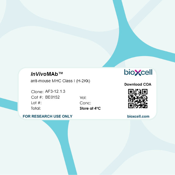
InVivoMAb anti-mouse MHC Class I (H-2Kk)
Product Description
Specifications
| Isotype | Mouse IgG1 |
|---|---|
| Recommended Isotype Control(s) | InVivoMAb mouse IgG1 isotype control, unknown specificity |
| Recommended Dilution Buffer | InVivoPure pH 7.0 Dilution Buffer |
| Conjugation | This product is unconjugated. Conjugation is available via our Antibody Conjugation Services. |
| Immunogen | A/J mouse spleen cells |
| Reported Applications | in vivo administration |
| Formulation |
PBS, pH 7.0 Contains no stabilizers or preservatives |
| Endotoxin |
≤1EU/mg (≤0.001EU/μg) Determined by LAL assay |
| Purity |
≥95% Determined by SDS-PAGE |
| Sterility | 0.2 µm filtration |
| Production | Purified from cell culture supernatant in an animal-free facility |
| Purification | Protein G |
| RRID | AB_10949012 |
| Molecular Weight | 150 kDa |
| Storage | The antibody solution should be stored at the stock concentration at 4°C. Do not freeze. |
| Need a Custom Formulation? | See All Antibody Customization Options |
Application References
in vivo administration
in vivo administration
Takenaka, M., et al (2012). "Complement activation is not required for obliterative airway disease induced by antibodies to major histocompatibility complex class I: Implications for chronic lung rejection" J Heart Lung Transplant 31(11): 1214-1222.
PubMed
BACKGROUND: The role of non-complement activating antibodies (ncAbs) to mismatched donor human leukocyte antigen (HLA) in the pathogenesis of chronic lung rejection is not known. We used a murine model of obliterative airway disease (OAD) induced by Abs to major histocompatibility major histocompatibility complex (MHC) class I and serum from donor-specific Abs developed in human lung transplant (LTx) recipients to test the role of ncAbs in the development of OAD and bronchiolitis obliterans syndrome (BOS). METHODS: Anti-MHC ncAbs were administered intrabronchially in B.10 mice or in C3 knockout (C3KO) mice. Lungs were analyzed by histopathology. Lymphocytes secreting interleukin (IL)-17, interferon-gamma, or IL-10 to collagen V and K-alpha1 tubulin (Kalpha1T) were enumerated by enzyme-linked immunospot assay. Serum antibodies to collagen V and Kalpha1T were determined by enzyme-linked immunosorbent assay. Cytokine and growth factor expression in lungs was determined by real-time polymerase chain reaction. Donor-specific Abs from patients with BOS and control BOS-negative LTx recipients were analyzed by C1q assay. RESULTS: Administration of ncAbs in B.10 mice or C3KO resulted in OAD lesions. There were significant increases in IL-17- and interferon-gamma-secreting cells to collagen V and Kalpha1T, along with serum Abs to these antigens. There was also augmented expression of monocyte chemotactic protein-1, IL-6, IL-1beta, vascular endothelial growth factor, transforming growth factor-beta, and fibroblastic growth factor in mice administered ncAbs by Day 3. Among 5 LTx recipients with BOS, only 1 had C1q binding donor-specific Abs. CONCLUSION: Complement activation by Abs to MHC class I is not required for development of OAD and human BOS. Therefore, anti-MHC binding to epithelial and endothelial cells can directly activate pro-fibrotic and pro-inflammatory cascades leading to immune response to self-antigens and chronic rejection.
in vivo administration
Hirohashi, T., et al (2010). "Complement independent antibody-mediated endarteritis and transplant arteriopathy in mice" Am J Transplant 10(3): 510-517.
PubMed
Complement fixation, as evidenced by C4d in the microvasculature, is a widely accepted criterion of antibody-mediated rejection. Complement fixation has been shown to be essential in acute antibody-mediated rejection, but its role in chronic rejection has not been addressed. Previous studies showed that passive transfer of complement fixing monoclonal IgG2a anti-H-2Kk into B6.RAG1-/- KO recipients of B10.BR hearts led to progressive chronic transplant arteriopathy (CTA) over 14-28 days, accompanied by C4d deposition. The present studies were designed to test whether complement was required for these lesions. We report that a noncomplement fixing donor-specific alloantibody (DSA, monoclonal IgG1 anti-H-2Kk) injected into B6.RAG1-/- KO recipients of B10.BR hearts also promotes CTA, without C4d deposition. Furthermore, a passive transfer of DSA (monoclonal IgG2a anti-H-2Kk) initiated endarteritis followed by CTA in B6.RAG1-/- mice genetically deficient in the third component of complement (RAG1-/-C3-/-). These studies indicate that antibody to class I MHC antigens can trigger chronic arterial lesions in vivo without complement participation, in contrast to acute antibody-mediated rejection. This pathway may be relevant to C4d-negative chronic rejection sometimes observed in patients with DSA, and argues that lack of C4d deposition does not exclude antibody-mediated chronic rejection.
in vivo administration
in vivo administration
Takenaka, M., et al (2012). "Complement activation is not required for obliterative airway disease induced by antibodies to major histocompatibility complex class I: Implications for chronic lung rejection" J Heart Lung Transplant 31(11): 1214-1222.
PubMed
BACKGROUND: The role of non-complement activating antibodies (ncAbs) to mismatched donor human leukocyte antigen (HLA) in the pathogenesis of chronic lung rejection is not known. We used a murine model of obliterative airway disease (OAD) induced by Abs to major histocompatibility major histocompatibility complex (MHC) class I and serum from donor-specific Abs developed in human lung transplant (LTx) recipients to test the role of ncAbs in the development of OAD and bronchiolitis obliterans syndrome (BOS). METHODS: Anti-MHC ncAbs were administered intrabronchially in B.10 mice or in C3 knockout (C3KO) mice. Lungs were analyzed by histopathology. Lymphocytes secreting interleukin (IL)-17, interferon-gamma, or IL-10 to collagen V and K-alpha1 tubulin (Kalpha1T) were enumerated by enzyme-linked immunospot assay. Serum antibodies to collagen V and Kalpha1T were determined by enzyme-linked immunosorbent assay. Cytokine and growth factor expression in lungs was determined by real-time polymerase chain reaction. Donor-specific Abs from patients with BOS and control BOS-negative LTx recipients were analyzed by C1q assay. RESULTS: Administration of ncAbs in B.10 mice or C3KO resulted in OAD lesions. There were significant increases in IL-17- and interferon-gamma-secreting cells to collagen V and Kalpha1T, along with serum Abs to these antigens. There was also augmented expression of monocyte chemotactic protein-1, IL-6, IL-1beta, vascular endothelial growth factor, transforming growth factor-beta, and fibroblastic growth factor in mice administered ncAbs by Day 3. Among 5 LTx recipients with BOS, only 1 had C1q binding donor-specific Abs. CONCLUSION: Complement activation by Abs to MHC class I is not required for development of OAD and human BOS. Therefore, anti-MHC binding to epithelial and endothelial cells can directly activate pro-fibrotic and pro-inflammatory cascades leading to immune response to self-antigens and chronic rejection.
in vivo administration
Hirohashi, T., et al (2010). "Complement independent antibody-mediated endarteritis and transplant arteriopathy in mice" Am J Transplant 10(3): 510-517.
PubMed
Complement fixation, as evidenced by C4d in the microvasculature, is a widely accepted criterion of antibody-mediated rejection. Complement fixation has been shown to be essential in acute antibody-mediated rejection, but its role in chronic rejection has not been addressed. Previous studies showed that passive transfer of complement fixing monoclonal IgG2a anti-H-2Kk into B6.RAG1-/- KO recipients of B10.BR hearts led to progressive chronic transplant arteriopathy (CTA) over 14-28 days, accompanied by C4d deposition. The present studies were designed to test whether complement was required for these lesions. We report that a noncomplement fixing donor-specific alloantibody (DSA, monoclonal IgG1 anti-H-2Kk) injected into B6.RAG1-/- KO recipients of B10.BR hearts also promotes CTA, without C4d deposition. Furthermore, a passive transfer of DSA (monoclonal IgG2a anti-H-2Kk) initiated endarteritis followed by CTA in B6.RAG1-/- mice genetically deficient in the third component of complement (RAG1-/-C3-/-). These studies indicate that antibody to class I MHC antigens can trigger chronic arterial lesions in vivo without complement participation, in contrast to acute antibody-mediated rejection. This pathway may be relevant to C4d-negative chronic rejection sometimes observed in patients with DSA, and argues that lack of C4d deposition does not exclude antibody-mediated chronic rejection.
in vivo administration
Takenaka, M., et al (2012). "Complement activation is not required for obliterative airway disease induced by antibodies to major histocompatibility complex class I: Implications for chronic lung rejection" J Heart Lung Transplant 31(11): 1214-1222.
PubMed
BACKGROUND: The role of non-complement activating antibodies (ncAbs) to mismatched donor human leukocyte antigen (HLA) in the pathogenesis of chronic lung rejection is not known. We used a murine model of obliterative airway disease (OAD) induced by Abs to major histocompatibility major histocompatibility complex (MHC) class I and serum from donor-specific Abs developed in human lung transplant (LTx) recipients to test the role of ncAbs in the development of OAD and bronchiolitis obliterans syndrome (BOS). METHODS: Anti-MHC ncAbs were administered intrabronchially in B.10 mice or in C3 knockout (C3KO) mice. Lungs were analyzed by histopathology. Lymphocytes secreting interleukin (IL)-17, interferon-gamma, or IL-10 to collagen V and K-alpha1 tubulin (Kalpha1T) were enumerated by enzyme-linked immunospot assay. Serum antibodies to collagen V and Kalpha1T were determined by enzyme-linked immunosorbent assay. Cytokine and growth factor expression in lungs was determined by real-time polymerase chain reaction. Donor-specific Abs from patients with BOS and control BOS-negative LTx recipients were analyzed by C1q assay. RESULTS: Administration of ncAbs in B.10 mice or C3KO resulted in OAD lesions. There were significant increases in IL-17- and interferon-gamma-secreting cells to collagen V and Kalpha1T, along with serum Abs to these antigens. There was also augmented expression of monocyte chemotactic protein-1, IL-6, IL-1beta, vascular endothelial growth factor, transforming growth factor-beta, and fibroblastic growth factor in mice administered ncAbs by Day 3. Among 5 LTx recipients with BOS, only 1 had C1q binding donor-specific Abs. CONCLUSION: Complement activation by Abs to MHC class I is not required for development of OAD and human BOS. Therefore, anti-MHC binding to epithelial and endothelial cells can directly activate pro-fibrotic and pro-inflammatory cascades leading to immune response to self-antigens and chronic rejection.
in vivo administration
Hirohashi, T., et al (2010). "Complement independent antibody-mediated endarteritis and transplant arteriopathy in mice" Am J Transplant 10(3): 510-517.
PubMed
Complement fixation, as evidenced by C4d in the microvasculature, is a widely accepted criterion of antibody-mediated rejection. Complement fixation has been shown to be essential in acute antibody-mediated rejection, but its role in chronic rejection has not been addressed. Previous studies showed that passive transfer of complement fixing monoclonal IgG2a anti-H-2Kk into B6.RAG1-/- KO recipients of B10.BR hearts led to progressive chronic transplant arteriopathy (CTA) over 14-28 days, accompanied by C4d deposition. The present studies were designed to test whether complement was required for these lesions. We report that a noncomplement fixing donor-specific alloantibody (DSA, monoclonal IgG1 anti-H-2Kk) injected into B6.RAG1-/- KO recipients of B10.BR hearts also promotes CTA, without C4d deposition. Furthermore, a passive transfer of DSA (monoclonal IgG2a anti-H-2Kk) initiated endarteritis followed by CTA in B6.RAG1-/- mice genetically deficient in the third component of complement (RAG1-/-C3-/-). These studies indicate that antibody to class I MHC antigens can trigger chronic arterial lesions in vivo without complement participation, in contrast to acute antibody-mediated rejection. This pathway may be relevant to C4d-negative chronic rejection sometimes observed in patients with DSA, and argues that lack of C4d deposition does not exclude antibody-mediated chronic rejection.