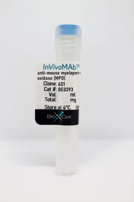
InVivoMAb anti-mouse myeloperoxidase (MPO)
Product Details
The 6D1 monoclonal antibody reacts with mouse myeloperoxidase (MPO). MPO is a peroxidase enzyme and a member of the subfamily of peroxidases. It is expressed primarily by neutrophils, but also by monocytes, macrophages, and some types of leukemic cells. MPO is a lysosomal protein stored in azurophilic granules of the neutrophil and released into the extracellular space during degranulation. MPO catalyzes the hydrogen peroxide dependent formation of hypochlorous acid and other reactive species. It is used by neutrophils to kill bacteria and other pathogens. Antibodies against MPO have been implicated in various types of vasculitis. Recent studies have reported an association between elevated myeloperoxidase levels and the severity of coronary artery disease. The 6D1 antibody is useful for inducing anti-neutrophil associated antibody (ANCA) mediated vasculitis in mice and is usually used in combination with the anti-mouse MPO 6G4 antibody for this purpose.Specifications
| Isotype | Mouse IgG2b, κ |
|---|---|
| Recommended Isotype Control(s) | InVivoMAb mouse IgG2b isotype control, unknown specificity |
| Recommended Dilution Buffer | InVivoPure pH 7.0 Dilution Buffer |
| Immunogen | Murine MPO |
| Reported Applications |
in vivo administration in vitro administration Immunofluorescence |
| Formulation |
PBS, pH 7.0 Contains no stabilizers or preservatives |
| Endotoxin |
<2EU/mg (<0.002EU/μg) Determined by LAL gel clotting assay |
| Sterility | 0.2 µm filtration |
| Purification | Protein A |
| Molecular Weight | 150 kDa |
| Storage | The antibody solution should be stored at the stock concentration at 4°C. Do not freeze. |
Recommended Products
in vivo administration
Fayçal CA, Oszwald A, Feilen T, Cosenza-Contreras M, Schilling O, Loustau T, Steinbach F, Schachner H, Langer B, Heeringa P, Rees AJ, Orend G, Kain R. (2022). "An adapted passive model of anti-MPO dependent crescentic glomerulonephritis reveals matrix dysregulation and is amenable to modulation by CXCR4 inhibition" Matrix Biol 106:12-33. PubMed
Anti-neutrophil cytoplasmic antibody (ANCA)-associated vasculitides (AAV) are severe inflammatory disorders that often involve focal necrotizing glomerulonephritis (FNGN) and consequent glomerular scarring, interstitial fibrosis, and chronic kidney disease. Robust murine models of scarring in FNGN that may help to further our understanding of deleterious processes are still lacking. Here, we present a murine model of severe FNGN based on combined administration of antibodies against the glomerular basement membrane (GBM) and myeloperoxidase (MPO), and bacterial lipopolysaccharides (LPS), that recapitulates acute injury and was adapted to investigate subsequent glomerular and interstitial scarring. Hematuria without involvement of other organs occurs consistently and rapidly, glomerular necrosis and crescent formation are evident at 12 days, and consequent glomerular and interstitial scarring at 29 days after initial treatment. Using mass-spectrometric proteome analysis, we provide a detailed overview of matrisomal and cellular changes in our model. We observed increased expression of the matrisome including collagens, fibronectin, tenascin-C, in accordance with human AAV as deduced from analysis of gene expression microarrays and tissue staining. Moreover, we observed tissue infiltration by neutrophils, macrophages, T cells and myofibroblasts upon injury. Experimental inhibition of CXCR4 using AMD3100 led to a sustained histological presence of fibrin extravasate, reduced chemokine expression and leukocyte activation, but did not markedly affect ECM composition. Altogether, we demonstrate an adapted FNGN model that enables the study of matrisomal changes both in disease and upon intervention, as exemplified via CXCR4 inhibition.
in vivo administration, in vitro administration, Immunofluorescence
Kessler N, Viehmann SF, Krollmann C, Mai K, Kirschner KM, Luksch H, Kotagiri P, Böhner AMC, Huugen D, de Oliveira Mann CC, Otten S, Weiss SAI, Zillinger T, Dobrikova K, Jenne DE, Behrendt R, Ablasser A, Bartok E, Hartmann G, Hopfner KP, Lyons PA, Boor P, Rösen-Wolff A, Teichmann LL, Heeringa P, Kurts C, Garbi N. (2022). "Monocyte-derived macrophages aggravate pulmonary vasculitis via cGAS/STING/IFN-mediated nucleic acid sensing" J Exp Med 219(10):e20220759. PubMed
Autoimmune vasculitis is a group of life-threatening diseases, whose underlying pathogenic mechanisms are incompletely understood, hampering development of targeted therapies. Here, we demonstrate that patients suffering from anti-neutrophil cytoplasmic antibodies (ANCA)-associated vasculitis (AAV) showed increased levels of cGAMP and enhanced IFN-I signature. To identify disease mechanisms and potential therapeutic targets, we developed a mouse model for pulmonary AAV that mimics severe disease in patients. Immunogenic DNA accumulated during disease onset, triggering cGAS/STING/IRF3-dependent IFN-I release that promoted endothelial damage, pulmonary hemorrhages, and lung dysfunction. Macrophage subsets played dichotomic roles in disease. While recruited monocyte-derived macrophages were major disease drivers by producing most IFN-β, resident alveolar macrophages contributed to tissue homeostasis by clearing red blood cells and limiting infiltration of IFN-β-producing macrophages. Moreover, pharmacological inhibition of STING, IFNAR-I, or its downstream JAK/STAT signaling reduced disease severity and accelerated recovery. Our study unveils the importance of STING/IFN-I axis in promoting pulmonary AAV progression and identifies cellular and molecular targets to ameliorate disease outcomes.


