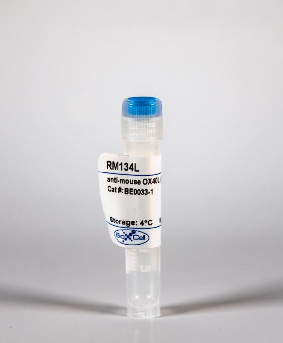
InVivoMAb anti-mouse OX40L (CD134L)
Product Details
The RM134L monoclonal antibody reacts with mouse OX-40L also known as CD134L. OX-40L is a 35 kDa member of the TNF superfamily that is expressed on activated B cells and antigen presenting cells. OX40L is the ligand for OX-40 (CD134). OX-40 signaling regulates both CD4 and CD8 T cell clonal expansion. It provides a costimulatory signal to an antigen-reacting naive T cells to prolong proliferation, as well as augment the production of several cytokines including IL-2. In vivo treatment with the RM134L antibody has been shown to inhibit the poly(I:C)/CD40 stimulated proliferation of CD4 T cells.Specifications
| Isotype | Rat IgG2b, κ |
|---|---|
| Recommended Isotype Control(s) | InVivoMAb rat IgG2b isotype control, anti-keyhole limpet hemocyanin |
| Recommended Dilution Buffer | InVivoPure pH 7.0 Dilution Buffer |
| Immunogen | Rat NRK-52E cells transfected with mouse OX40L |
| Reported Applications |
in vivo blocking of OX40/OX40L signaling in vitro OX40L neutralization |
| Formulation |
PBS, pH 7.0 Contains no stabilizers or preservatives |
| Endotoxin |
<2EU/mg (<0.002EU/μg) Determined by LAL gel clotting assay |
| Sterility | 0.2 μM filtered |
| Production | Purified from tissue culture supernatant in an animal free facility |
| Purification | Protein G |
| RRID | AB_1107594 |
| Molecular Weight | 150 kDa |
| Storage | The antibody solution should be stored at the stock concentration at 4°C. Do not freeze. |
Recommended Products
in vivo blocking of OX40/OX40L signaling
Gattringer, M., et al. (2015). "Anti-OX40L alone or in combination with anti-CD40L and CTLA4Ig does not inhibit the humoral and cellular response to a major grass pollen allergen" Clin Exp Allergy. doi : 10.1111/cea.12661. PubMed
BACKGROUND: IgE-mediated allergy is a common disease characterized by a harmful immune response towards otherwise harmless environmental antigens. Induction of specific immunological non-responsiveness towards allergens would be a desirable goal. Blockade of costimulatory pathways is a promising strategy to modulate the immune response in an antigen-specific manner. Recently OX40 (CD134) was identified as a costimulatory receptor important in Th2 mediated immune responses. Moreover, synergy between OX40 blockade and ‘conventional’ costimulation blockade (anti-CD40L, CTLA4Ig) was observed in models of alloimmunity. OBJECTIVE: We investigated the potential of interfering with OX40 alone or in combination with CD40/CD28 signals to influence the allergic immune response. METHODS: The OX40 pathway was investigated in an established murine model of IgE-mediated allergy where BALB/c mice are repeatedly immunized with the clinically relevant grass pollen allergen Phl p 5. Groups were treated with combinations of anti-OX40L, CTLA4Ig and anti-CD40L. In selected mice Tregs were depleted with anti-CD25. RESULTS: Blockade of OX40L alone at the time of first or second immunization did not modulate the allergic response on the humoral or effector cell levels but slightly on T cell responses. Administration of a combination of anti-CD40L/CTLA4Ig delayed the allergic immune response but antibody-production could not be inhibited after repeated immunization even though the allergen-specific T cell response was suppressed in the long run. Notably, additional blockade of OX40L had no detectable supplementary effect. Immunomodulation partly involved regulatory T cells as depletion of CD25+ cells led to restored T cell proliferation. CONCLUSIONS AND CLINICAL RELEVANCE: Collectively, our data provide evidence that the allergic immune response towards Phl p 5 is independent of OX40L, although reduction on T cell responses and slightly, on the asthmatic phenotype, were detectable. Besides, no relevant synergistic effect of OX40L blockade in addition to CD40L/CD28 blockade could be detected. Thus, the therapeutic potential of OX40L blockade for IgE-mediated allergy appears to be ineffective in this setting. This article is protected by copyright. All rights reserved.
in vivo blocking of OX40/OX40L signaling
Baeyens, A., et al. (2015). "Effector T cells boost regulatory T cell expansion by IL-2, TNF, OX40, and plasmacytoid dendritic cells depending on the immune context" J Immunol 194(3): 999-1010. PubMed
CD4(+)CD25(+)Foxp3(+) regulatory T (Treg) cells play a major role in peripheral tolerance. Multiple environmental factors and cell types affect their biology. Among them, activated effector CD4(+) T cells can boost Treg cell expansion through TNF or IL-2. In this study, we further characterized this effector T (Teff) cell-dependent Treg cell boost in vivo in mice. This phenomenon was observed when both Treg and Teff cells were activated by their cognate Ag, with the latter being the same or different. Also, when Treg cells highly proliferated on their own, there was no additional Treg cell boost by Teff cells. In a condition of low inflammation, the Teff cell-mediated Treg cell boost involved TNF, OX40L, and plasmacytoid dendritic cells, whereas in a condition of high inflammation, it involved TNF and IL-2. Thus, this feedback mechanism in which Treg cells are highly activated by their Teff cell counterparts depends on the immune context for its effectiveness and mechanism. This Teff cell-dependent Treg cell boost may be crucial to limit inflammatory and autoimmune responses.
in vivo blocking of OX40/OX40L signaling
Welten, S. P., et al. (2015). "The viral context instructs the redundancy of costimulatory pathways in driving CD8(+) T cell expansion" Elife 4. doi : 10.7554/eLife.07486. PubMed
Signals delivered by costimulatory molecules are implicated in driving T cell expansion. The requirements for these signals, however, vary from dispensable to essential in different infections. We examined the underlying mechanisms of this differential T cell costimulation dependence and found that the viral context determined the dependence on CD28/B7-mediated costimulation for expansion of naive and memory CD8(+) T cells, indicating that the requirement for costimulatory signals is not imprinted. Notably, related to the high-level costimulatory molecule expression induced by lymphocytic choriomeningitis virus (LCMV), CD28/B7-mediated costimulation was dispensable for accumulation of LCMV-specific CD8(+) T cells because of redundancy with the costimulatory pathways induced by TNF receptor family members (i.e., CD27, OX40, and 4-1BB). Type I IFN signaling in viral-specific CD8(+) T cells is slightly redundant with costimulatory signals. These results highlight that pathogen-specific conditions differentially and uniquely dictate the utilization of costimulatory pathways allowing shaping of effector and memory antigen-specific CD8(+) T cell responses.
in vivo blocking of OX40/OX40L signaling
Xin, L., et al. (2014). "Commensal microbes drive intestinal inflammation by IL-17-producing CD4+ T cells through ICOSL and OX40L costimulation in the absence of B7-1 and B7-2" Proc Natl Acad Sci U S A 111(29): 10672-10677. PubMed
The costimulatory B7-1 (CD80)/B7-2 (CD86) molecules, along with T-cell receptor stimulation, together facilitate T-cell activation. This explains why in vivo B7 costimulation neutralization efficiently silences a variety of human autoimmune disorders. Paradoxically, however, B7 blockade also potently moderates accumulation of immune-suppressive regulatory T cells (Tregs) essential for protection against multiorgan systemic autoimmunity. Here we show that B7 deprivation in mice overrides the necessity for Tregs in averting systemic autoimmunity and inflammation in extraintestinal tissues, whereas peripherally induced Tregs retained in the absence of B7 selectively mitigate intestinal inflammation caused by Th17 effector CD4(+) T cells. The need for additional immune suppression in the intestine reflects commensal microbe-driven T-cell activation through the accessory costimulation molecules ICOSL and OX40L. Eradication of commensal enteric bacteria mitigates intestinal inflammation and IL-17 production triggered by Treg depletion in B7-deficient mice, whereas re-establishing intestinal colonization with Candida albicans primes expansion of Th17 cells with commensal specificity. Thus, neutralizing B7 costimulation uncovers an essential role for Tregs in selectively averting intestinal inflammation by Th17 CD4(+) T cells with commensal microbe specificity.
in vitro OX40L neutralization
Mahmud, S. A., et al. (2014). "Costimulation via the tumor-necrosis factor receptor superfamily couples TCR signal strength to the thymic differentiation of regulatory T cells" Nat Immunol 15(5): 473-481. PubMed
Regulatory T cells (Treg cells) express members of the tumor-necrosis factor (TNF) receptor superfamily (TNFRSF), but the role of those receptors in the thymic development of Treg cells is undefined. We found here that Treg cell progenitors had high expression of the TNFRSF members GITR, OX40 and TNFR2. Expression of those receptors correlated directly with the signal strength of the T cell antigen receptor (TCR) and required the coreceptor CD28 and the kinase TAK1. The neutralization of ligands that are members of the TNF superfamily (TNFSF) diminished the development of Treg cells. Conversely, TNFRSF agonists enhanced the differentiation of Treg cell progenitors by augmenting responsiveness of the interleukin 2 receptor (IL-2R) and transcription factor STAT5. Costimulation with the ligand of GITR elicited dose-dependent enrichment for cells of lower TCR affinity in the Treg cell repertoire. In vivo, combined inhibition of GITR, OX40 and TNFR2 abrogated the development of Treg cells. Thus, expression of members of the TNFRSF on Treg cell progenitors translated strong TCR signals into molecular parameters that specifically promoted the development of Treg cells and shaped the Treg cell repertoire.
in vivo blocking of OX40/OX40L signaling
Hong, J., et al. (2013). "Islet allograft rejection in sensitized mice is refractory to control by combination therapy of immune-modulating agents" Transpl Immunol 28(2-3): 86-92. PubMed
Retransplantation is common in allogeneic islet transplantation, and therefore, memory responses in previously sensitized recipients present a distinct obstacle for successful islet transplantation. Given the difficulties in controlling memory responses contributing to allograft rejection, it is worth investigating the effects of new immune-modulating agents against islet allograft rejection in the sensitized recipients. In this study, we investigated immune-modulating agents including 5-azacytidine and IL-2/anti-IL-2 complex to ascertain their suppressive effects on memory responses. In suppression assays, rapamycin effectively suppressed the proliferation of memory T cells, whereas 5-azacytidine, a methylation inhibitor suppressed the survival and proliferation of memory T cells. Combination therapy of anti-CD40L, anti-OX40L, and rapamycin slightly prolonged BALB/c islet allograft survival in sensitized C57BL6 mice, and reduced intragraft infiltration of macrophages, T cells, and B cells. However, the addition of IL-2/anti-IL-2 complex, an inducer of regulatory T cells, did not exhibit additional suppression against rejection in sensitized mice. Although a combination of 5-azacytidine and rapamycin markedly suppressed islet allograft rejection in naive mice, it failed to achieve long-term graft survival even when combined with anti-CD40L and anti-OX40 in sensitized mice. In short, 5-azacytidine-based or IL-2/anti-IL-2 complex-based regimens can suppress islet allograft rejection in naive recipients, but fail to control islet allograft rejection in sensitized recipients.
in vivo blocking of OX40/OX40L signaling
Leyva-Castillo, J. M., et al. (2013). "Skin thymic stromal lymphopoietin initiates Th2 responses through an orchestrated immune cascade" Nat Commun 4: 2847. PubMed
Thymic stromal lymphopoietin (TSLP) has emerged as a key initiator in Th2 immune responses, but the TSLP-driven immune cascade leading to Th2 initiation remains to be delineated. Here, by dissecting the cellular network triggered by mouse skin TSLP in vivo, we uncover that TSLP-promoted IL-4 induction in CD4(+) T cells in skin-draining lymph nodes is driven by an orchestrated ‘DC-T-Baso-T’ cascade, which represents a sequential cooperation of dendritic cells (DCs), CD4(+) T cells and basophils. Moreover, we reveal that TSLP-activated DCs prime naive CD4(+) T cells to produce IL-3 via OX40L signalling and demonstrate that the OX40L-IL-3 axis has a critical role in mediating basophil recruitment, CD4(+) T-cell expansion and Th2 priming. These findings thus add novel insights into the cellular network and signal axis underlying the initiation of Th2 immune responses.
in vivo blocking of OX40/OX40L signaling
Sanchez, P. J. and R. M. Kedl. (2012). "An alternative signal 3: CD8(+) T cell memory independent of IL-12 and type I IFN is dependent on CD27/OX40 signaling" Vaccine 30(6): 1154-1161. PubMed
Type I IFN and IL-12 are well documented to serve as so called “signal 3” cytokines, capable of facilitating CD8(+) T cell proliferation, effector function and memory formation. While their ability to serve in this capacity is well established, to date, no non-cytokine signal 3 mediators have been clearly identified. We have established a vaccine model system in which the primary CD8(+) T cell response is independent of either IL-12 or type I IFN receptors, but dependent on CD27/CD70 interactions. We show here that primary and secondary CD8(+) T cell responses are generated in the combined deficiency of IFN and IL-12 signaling. In contrast, antigen specific CD8(+) T cell responses are compromised in the absence of the TNF receptors CD27 and OX40. These data indicate that CD27/OX40 can serve the central function as signal 3 mediators, independent of IFN or IL-12, for the generation of CD8(+) T cell immune memory.
in vivo blocking of OX40/OX40L signaling
Kurche, J. S., et al. (2010). "Comparison of OX40 ligand and CD70 in the promotion of CD4+ T cell responses" J Immunol 185(4): 2106-2115. PubMed
The TNF superfamily members CD70 and OX40 ligand (OX40L) were reported to be important for CD4(+) T cell expansion and differentiation. However, the relative contribution of these costimulatory signals in driving CD4(+) T cell responses has not been addressed. In this study, we found that OX40L is a more important determinant than CD70 of the primary CD4(+) T cell response to multiple immunization regimens. Despite the ability of a combined TLR and CD40 agonist (TLR/CD40) stimulus to provoke appreciable expression of CD70 and OX40L on CD8(+) dendritic cells, resulting CD4(+) T cell responses were substantially reduced by Ab blockade of OX40L and, to a lesser degree, CD70. In contrast, the CD8(+) T cell responses to combined TLR/CD40 immunization were exclusively dependent on CD70. These requirements for CD4(+) and CD8(+) T cell activation were not limited to the use of combined TLR/CD40 immunization, because vaccinia virus challenge elicited primarily OX40L-dependent CD4 responses and exclusively CD70-dependent CD8(+) T cell responses. Attenuation of CD4(+) T cell priming induced by OX40L blockade was independent of signaling through the IL-12R, but it was reduced further by coblockade of CD70. Thus, costimulation by CD70 or OX40L seems to be necessary for primary CD4(+) T cell responses to multiple forms of immunization, and each may make independent contributions to CD4(+) T cell priming.
in vivo blocking of OX40/OX40L signaling
Vu, M. D., et al. (2006). "Critical, but conditional, role of OX40 in memory T cell-mediated rejection" J Immunol 176(3): 1394-1401. PubMed
100 days. In contrast, blocking the ICOS/ICOS ligand and the 4-1BB/4-1BBL pathways alone or combined with CD28/CD154 blockade had no effect in preventing skin allograft rejection. OX40 blockade did not affect the homeostatic proliferation of T cells in vivo, but markedly inhibited the effector functions of memory T cells. Our data demonstrate that memory T cells resisting to CD28/CD154 blockade in transplant rejection are sensitive to OX40 blockade and suggest that OX40 is a key therapeutic target in memory T cell-mediated rejection.”}” data-sheets-userformat=”{“2″:14851,”3”:{“1″:0},”4”:{“1″:2,”2″:16777215},”12″:0,”14”:{“1″:2,”2″:1521491},”15″:”Roboto, sans-serif”,”16″:12}”>Memory T cells can be a significant barrier to the induction of transplant tolerance. However, the molecular pathways that can regulate memory T cell-mediated rejection are poorly defined. In the present study we tested the hypothesis that the novel alternative costimulatory molecules (i.e., ICOS, 4-1BB, OX40, or CD30) may play a critical role in memory T cell activation and memory T cell-mediated rejection. We found that memory T cells, generated by either homeostatic proliferation or donor Ag priming, induced prompt skin allograft rejection regardless of CD28/CD154 blockade. Phenotypic analysis showed that, in contrast to naive T cells, such memory T cells expressed high levels of OX40, 4-1BB, and ICOS on the cell surface. In a skin transplant model in which rejection was mediated by memory T cells, blocking the OX40/OX40 ligand pathway alone did not prolong the skin allograft survival, but blocking OX40 costimulation in combination with CD28/CD154 blockade induced long-term skin allograft survival, and 40% of the recipients accepted their skin allograft for >100 days. In contrast, blocking the ICOS/ICOS ligand and the 4-1BB/4-1BBL pathways alone or combined with CD28/CD154 blockade had no effect in preventing skin allograft rejection. OX40 blockade did not affect the homeostatic proliferation of T cells in vivo, but markedly inhibited the effector functions of memory T cells. Our data demonstrate that memory T cells resisting to CD28/CD154 blockade in transplant rejection are sensitive to OX40 blockade and suggest that OX40 is a key therapeutic target in memory T cell-mediated rejection.


