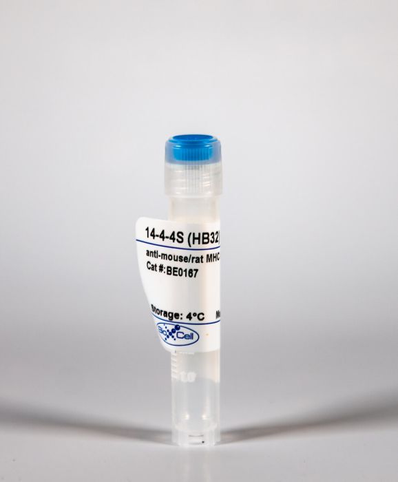
InVivoMAb anti-mouse/rat MHC Class II (I-Ek/RT1-D)
Product Details
The 14-4-4S monoclonal antibody reacts with mouse MHC Class II alloantigen I-Ek and the rat MHC class II alloantigen RT1D. These MHC class II molecules are expressed primarily on the surface of B lymphocytes, macrophages, dendritic cells and other antigen presenting cells as well as a subset of T cells from H-2k bearing mice. These MHC molecules play a role in antigen presentation to T cells. The 14-4-4S antibody has been reported to block antigen presentation and induce differentiation of mouse cells expressing I-Ek.Specifications
| Isotype | Mouse IgG2a, κ |
|---|---|
| Recommended Isotype Control(s) | InVivoMAb mouse IgG2a isotype control, unknown specificity |
| Recommended Dilution Buffer | InVivoPure pH 7.0 Dilution Buffer |
| Immunogen | C3H mouse skin graft and spleen cells |
| Reported Applications |
in vivo blocking of antigen presentation Flow cytometry |
| Formulation |
PBS, pH 7.0 Contains no stabilizers or preservatives |
| Endotoxin |
<2EU/mg (<0.002EU/μg) Determined by LAL gel clotting assay |
| Sterility | 0.2 μM filtered |
| Production | Purified from tissue culture supernatant in an animal free facility |
| Purification | Protein G |
| RRID | AB_10950190 |
| Molecular Weight | 150 kDa |
| Storage | The antibody solution should be stored at the stock concentration at 4°C. Do not freeze. |
Recommended Products
Flow Cytometry, in vivo blocking of antigen presentation
Positional identification of RT1-B (HLA-DQ) as susceptibility locus for autoimmune arthritis PubMed
Rheumatoid arthritis (RA) is associated with amino acid variants in multiple MHC molecules. The association to MHC class II (MHC-II) has been studied in several animal models of RA. In most cases these models depend on T cells restricted to a single immunodominant peptide of the immunizing Ag, which does not resemble the autoreactive T cells in RA. An exception is pristane-induced arthritis (PIA) in the rat where polyclonal T cells induce chronic arthritis after being primed against endogenous Ags. In this study, we used a mixed genetic and functional approach to show that RT1-Ba and RT1-Bb (RT1-B locus), the rat orthologs of HLA-DQA and HLA-DQB, determine the onset and severity of PIA. We isolated a 0.2-Mb interval within the MHC-II locus of three MHC-congenic strains, of which two were protected from severe PIA. Comparison of sequence and expression variation, as well as in vivo blocking of RT1-B and RT1-D (HLA-DR), showed that arthritis in these strains is regulated by coding polymorphisms in the RT1-B genes. Motif prediction based on MHC-II eluted peptides and structural homology modeling suggested that variants in the RT1-B P1 pocket, which likely affect the editing capacity by RT1-DM, are important for the development of PIA.


