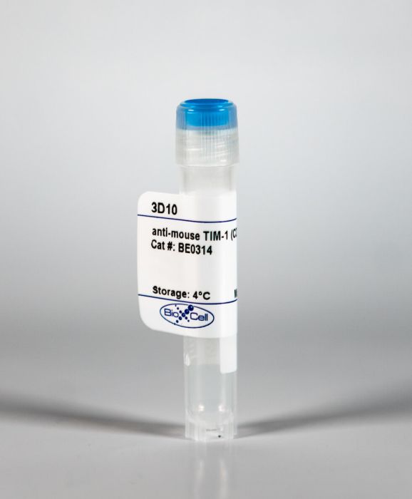
InVivoMAb anti-mouse TIM-1 (CD365)
Product Details
The 3D10 monoclonal antibody reacts with mouse T cell immunoglobulin and mucin domain 1 (TIM-1) also known as CD365. TIM-1 is a type I cell-surface glycoprotein and member of the Ig superfamily. TIM-1 is preferentially expressed on TH2 cells and has been identified as a stimulatory molecule for T cell activation. The TIM gene family, plays critical roles in regulating the immune response to viral infection. TIM-1 is also involved in allergic responses, asthma, and transplant tolerance. The 3D10 antibody has been shown to block TIM-1 in vivo and enhance atherosclerosis in mice studies.Specifications
| Isotype | Rat IgG1, κ |
|---|---|
| Recommended Isotype Control(s) | InVivoMAb rat IgG1 isotype control, anti-horseradish peroxidase |
| Recommended Dilution Buffer | InVivoPure pH 7.0 Dilution Buffer |
| Immunogen | Mouse TIM-1 (signal and IgV domains)/mouse IgG2a Fc fusion protein |
| Reported Applications |
in vivo TIM-1 blockade in vitro TIM-1 blockade |
| Formulation |
PBS, pH 7.0 Contains no stabilizers or preservatives |
| Endotoxin |
<2EU/mg (<0.002EU/μg) Determined by LAL gel clotting assay |
| Sterility | 0.2 μM filtered |
| Production | Purified from tissue culture supernatant in an animal free facility |
| Purification | Protein G |
| RRID | AB_2754552 |
| Molecular Weight | 150 kDa |
| Storage | The antibody solution should be stored at the stock concentration at 4°C. Do not freeze. |
Recommended Products
Functional assays, in vitro TIM-1 blockade, in vivo TIM-1 blockade
Apoptotic cells activate NKT cells through T cell Ig-like mucin-like-1 resulting in airway hyperreactivity PubMed
T cell Ig-like mucin-like-1 (TIM-1) is an important asthma susceptibility gene, but the immunological mechanisms by which TIM-1 functions remain uncertain. TIM-1 is also a receptor for phosphatidylserine (PtdSer), an important marker of cells undergoing programmed cell death, or apoptosis. We now demonstrate that NKT cells constitutively express TIM-1 and become activated by apoptotic cells expressing PtdSer. TIM-1 recognition of PtdSer induced NKT cell activation, proliferation, and cytokine production. Moreover, the induction of apoptosis in airway epithelial cells activated pulmonary NKT cells and unexpectedly resulted in airway hyperreactivity, a cardinal feature of asthma, in an NKT cell-dependent and TIM-1-dependent fashion. These results suggest that TIM-1 serves as a pattern recognition receptor on NKT cells that senses PtdSer on apoptotic cells as a damage-associated molecular pattern. Furthermore, these results provide evidence for a novel innate pathway that results in airway hyperreactivity and may help to explain how TIM-1 and NKT cells regulate asthma.
in vivo TIM-1 blockade
T-cell immunoglobulin and mucin domain 1 deficiency eliminates airway hyperreactivity triggered by the recognition of airway cell death PubMed
BACKGROUND: Studies of asthma have been limited by a poor understanding of how nonallergic environmental exposures, such as air pollution and infection, are translated in the lung into inflammation and wheezing. OBJECTIVE: Our goal was to understand the mechanism of nonallergic asthma that leads to airway hyperreactivity (AHR), a cardinal feature of asthma independent of adaptive immunity. METHOD: We examined mouse models of experimental asthma in which AHR was induced by respiratory syncytial virus infection or ozone exposure using mice deficient in T-cell immunoglobulin and mucin domain 1 (TIM1/HAVCR1), an important asthma susceptibility gene. RESULTS: TIM1(-/-) mice did not have airways disease when infected with RSV or when repeatedly exposed to ozone, a major component of air pollution. On the other hand, the TIM1(-/-) mice had allergen-induced experimental asthma, as previously shown. The RSV- and ozone-induced pathways were blocked by treatment with caspase inhibitors, indicating an absolute requirement for programmed cell death and apoptosis. TIM-1-expressing, but not TIM-1-deficient, natural killer T cells responded to apoptotic airway epithelial cells by secreting cytokines, which mediated the development of AHR. CONCLUSION: We defined a novel pathway in which TIM-1, a receptor for phosphatidylserine expressed by apoptotic cells, drives the development of asthma by sensing and responding to injured and apoptotic airway epithelial cells.
in vivo TIM-1 blockade
Blockade of Tim-1 and Tim-4 Enhances Atherosclerosis in Low-Density Lipoprotein Receptor-Deficient Mice PubMed
OBJECTIVE: T cell immunoglobulin and mucin domain (Tim) proteins are expressed by numerous immune cells, recognize phosphatidylserine on apoptotic cells, and function as costimulators or coinhibitors. Tim-1 is expressed by activated T cells but is also found on dendritic cells and B cells. Tim-4, present on macrophages and dendritic cells, plays a critical role in apoptotic cell clearance, regulates the number of phosphatidylserine-expressing activated T cells, and is genetically associated with low low-density lipoprotein and triglyceride levels. Because these functions of Tim-1 and Tim-4 could affect atherosclerosis, their modulation has potential therapeutic value in cardiovascular disease. APPROACH AND RESULTS: ldlr(-/-) mice were fed a high-fat diet for 4 weeks while being treated with control (rat immunoglobulin G1) or anti-Tim-1 (3D10) or -Tim-4 (21H12) monoclonal antibodies that block phosphatidylserine recognition and phagocytosis. Both anti-Tim-1 and anti-Tim-4 treatments enhanced atherosclerosis by 45% compared with controls by impairment of efferocytosis and increasing aortic CD4(+)T cells. Consistently, anti-Tim-4-treated mice showed increased percentages of activated T cells and late apoptotic cells in the circulation. Moreover, in vitro blockade of Tim-4 inhibited efferocytosis of oxidized low-density lipoprotein-induced apoptotic macrophages. Although anti-Tim-4 treatment increased T helper cell (Th)1 and Th2 responses, anti-Tim-1 induced Th2 responses but dramatically reduced the percentage of regulatory T cells. Finally, combined blockade of Tim-1 and Tim-4 increased atherosclerotic lesion size by 59%. CONCLUSIONS: Blockade of Tim-4 aggravates atherosclerosis likely by prevention of phagocytosis of phosphatidylserine-expressing apoptotic cells and activated T cells by Tim-4-expressing cells, whereas Tim-1-associated effects on atherosclerosis are related to changes in Th1/Th2 balance and reduced circulating regulatory T cells.


