InVivoMAb anti-rat CD4
Product Description
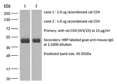
Specifications
| Isotype | Mouse IgG1, κ |
|---|---|
| Recommended Isotype Control(s) | InVivoMAb rat IgG2b isotype control, anti-keyhole limpet hemocyanin |
| Recommended Dilution Buffer | InVivoPure pH 7.0 Dilution Buffer |
| Reported Applications |
in vivo down-regulation of surface CD4 in vitro neutralization of CD4 Flow cytometry Immunohistochemistry (paraffin) Immunohistochemistry (frozen) |
| Formulation |
PBS, pH 7.0 Contains no stabilizers or preservatives |
| Endotoxin |
≤1EU/mg (≤0.001EU/μg) Determined by LAL assay |
| Purity |
≥95% Determined by SDS-PAGE |
| Sterility | 0.2 µm filtration |
| Production | Purified from cell culture supernatant in an animal-free facility |
| Purification | Protein G |
| Molecular Weight | 150 kDa |
| Storage | The antibody solution should be stored at the stock concentration at 4°C. Do not freeze. |
| Need a Custom Formulation? | See All Antibody Customization Options |
Application References
-
Mannie MD, White GA, Nardella JP, Davidian DK, Arnold PY (1998). "Partial agonism elicits an enduring phase of T-cell-medicated antigen presentation" Cell Immunol 186(2):83-93.
PubMed
Previous studies have shown that the anti-CD4 mAb W3/25 strongly enhances T cell APC (T-APC) activity. In this study, single positive CD4+ and double negative (DN) (CD4-CD8-) T-helper cells specific for the 55-69 or 72-86 sequence of guinea pig (GP) myelin basic protein (GPMBP) were used to study CD4 regulation of T-APC activity. Clones were cultured with irradiated SPL and GPMBP or rat (R) MBP for 2-3 days, were propagated in IL-2 for another 1-3 days, were irradiated, and were used as T-APC. DN T cells specific for GP55-69 effectively presented GPMBP and were superior APC compared to other CD4+ T cells for presentation of this antigen. In contrast, DN T cells specific for the dominant encephalitogenic 72-86 determinant did not effectively present the agonist GPMBP but potently presented the partial agonist RMBP. The heightened APC activity of DN T cells reflected the lack of CD4 because the anti-CD4 mAb W3/25 promoted T-APC activity of CD4+ T cells to those levels expressed by DN T cells. Overall, T cells with potent reactivity to GPMBP or RMBP were subsequently unable to present that antigen, whereas T cells exhibiting partial or low antigen reactivities were highly effective APC for presentation of that antigen. The unrelated antigen conalbumin was presented by MBP-specific clones only when added to culture with a specific partial agonist. Together, these data indicate that partially agonistic MHC ligands promote prolonged expression of T-APC activity and that DN T cells may be specialized to mediate postactivational antigen presentation.
-
Mannie MD, Rosser JM, White GA (1995). "Autologous rat myelin basic protein is a partial agonist that is converted into a full antagonist upon blockade of CD4. Evidence for the integration of efficacious and nonefficacious signals during T cell antig
PubMed
A central question of TCR function is based on the observation that some MHC ligands may bind TCR without stimulating biologic activity. To address the role of CD4 in this mechanism, we studied the interactions of the anti-CD4 mAb W3/25 with an encephalitogenic line of T helper cells. Proliferative responses to guinea pig (GP) myelin basic protein (GPMBP) were mediated by distinct W3/25-sensitive and W3/25-insensitive mechanisms whereas responses to autologous rat (R) MBP (RMBP) were exclusively mediated by W3/25-sensitive pathways. In assays of IL-2 production, RMBP was a partial agonist that stimulated an intermediate level of IL-2 production but antagonized high levels of GPMBP-stimulated IL-2 production to that intermediate level. In the presence of W3/25, RMBP lacked stimulatory activity but instead exhibited inhibitory activity that completely blocked GPMBP-stimulated proliferative responses. The inhibitory mechanism did not involve antigenic competition for MHC glycoproteins, a blockade of mitogenic signaling, or induction of high zone tolerance. Rather, the mechanism involved specific occupancy of TCR with antagonistic MHC ligands derived from the 72-86 region of RMBP. In proliferative assays, GPMBP was approximately 10-fold more active than RMBP. In the presence of W3/25 however, GPMBP-induced agonism and RMBP-induced antagonism exhibited overlapping dose-response curves. RMBP and W3/25 not only fully inhibited GPMBP-stimulated proliferation, this synergistic combination also elicited an extended phase of T cell energy. In conclusion, RMBP-derived MHC ligands occupy TCR to exhibit full efficacy for CD4-dependent signaling pathways while simultaneously lacking efficacy for W3/25-resistant signaling pathways. These data support an "integrative" model of T cell Ag recognition whereby MHC glycoproteins actively guide specific clustering of MHC-ligated TCR to enable quantitative comparisons of efficacious (nonself) and nonefficacious (self) signals by T cells.
-
Remuzzi G, Zoja C, Gagliardini E, Corna D, Abbate M, Benigni A (1999). "Combining an antiproteinuric approach with mycophenolate mofetil fully suppresses progressive nephropathy of experimental animals" J Am Soc Nephrol 10(7):1542-9.
PubMed
Chronic renal diseases progress to organ insufficiency, which may require replacement therapy within one to three decades even independently of the type of initial insults. In the majority of cases, the degrees of proteinuria and interstitial leukocyte infiltration and scarring are strictly correlated with the rate of disease progression. This study tests the hypothesis that excess intrarenal protein traffic may cause lymphocyte-dependent interstitial injury that, while not fully controlled by antiproteinuric therapy, can be further inhibited by concomitant immunosuppression. A primarily nonimmune model was used to reproduce progressive renal disease due to a critical loss of nephron mass. Angiotensin-converting enzyme (ACE) inhibitor limited proteinuria, interstitial inflammation, MHC class II antigen expression, and severe lesions. Combined treatment with ACE inhibitor and a specific antilymphocyte agent, mycophenolate mofetil, dramatically attenuated macrophage and T cell infiltration, MHC-class II overexpression, dendritic cells, and all manifestations of the disease. Evidence of lymphocyte-mediated renal injury in the setting of excess protein traffic provides the basis for combining ACE inhibition and immunosuppression to halt progression of proteinuric kidney disease and minimize the need for dialysis or transplantation.
-
Ysebaert DK, De Greef KE, Vercauteren SR, Ghielli M, Verpooten GA, Eyskens EJ, De Broe ME (2000). "Identification and kinetics of leukocytes after severe ischaemia/reperfusion renal injury" Nephrol Dial Transplant 15(10):1562-74.
PubMed
Background: Leukocyte adhesion/infiltration in response to renal ischaemia/reperfusion (I/R) injury is a well-known but poorly understood phenomenon. The identification, kinetics, and exact role of these inflammatory cells in I/R injury and regeneration are still matters of debate. Methods: Uninephrectomized rats were submitted to 60 min renal ischaemia by clamping of renal vessels. Results: Severe acute renal failure was observed, with maximum functional impairment on day 2. By 12 h after the ischaemic event, up to 80% of proximal tubular cells in the outer stripe of outer medulla (OSOM) were already severely damaged. Proliferation (proliferating cell nuclear antigen (PCNA) staining) started after 24 h, reaching maximum activity on day 3. Regeneration of tubular morphology started on the 3rd day, and after 10 days 50% of tubules had regenerated completely. Interstitial leukocytes (OX-1 immunohistochemical staining) were already prominent at day 1, thereafter gradually increasing with time. The so-called neutrophil-specific identification methods (myeloperoxidase (MPO), chloroacetate esterase, mAb HIS-48) proved to be non-specific, since they also stained for macrophages, as demonstrated by flow cytometry and the combination of these stainings with the macrophage-specific ED-1 staining. MPO activity was already significantly increased at 1 h post-I/R (439+/-34%, P<0.005), reaching its maximum activity after 12 h of I/R (1159+/-138%, P<0.0005), declining thereafter. On the other hand, neutrophil presence investigated by H&E staining revealed only a few neutrophils in glomeruli, medullary rays, and OSOM at 24 h after the ischaemic event (4.7+/-4.2 cells/mm(2) vs controls=2.3+/-2.0 cells/mm(2) (n.s.)), and remained unchanged over the next 10 days. In contrast, significant monocyte/macrophage adhesion/infiltration (ED-1 staining) occurred at the OSOM at 24 h post-ischaemia (at 24 h, 120+/-46 cells/mm(2) vs. sham=18+/-4 cells/mm(2) (P<0.05)), became prominent at day 5 (1034+/-161 cells/mm(2) vs sham=18+/-18 cells/mm(2) (P<0.05)), and almost disappeared after 10 days. CD4(+) cells (W3/25) gradually increased from day 5, reaching a maximum at day 10. A few CD8(+) cells (OX-8) were apparent from days 3 until 10, but no B-cells (OX-33) were observed. Conclusions: After severe warm I/R renal injury, a pronounced acute tubular necrosis occurs during the first 12-24 h in the absence of a marked cellular infiltrate, but with an important renal MPO activity, reflecting the activation of the adhering inflammatory cells (polymorphonuclear cells (PMNs) and mainly monocytes/macrophages). Only later at the time and site (OSOM) of regeneration a sequential accumulation of monocytes/macrophages and T cells becomes prominent, in contrast with the low number of neutrophils found in the kidney during the 10-day post-ischaemic period. The non-specificity of the so-called neutrophil-specific identification methods (MPO activity, naphthol AS-D chloroacetate esterase, or mAb HIS-48 staining), cross-reacting with monocytes/macrophages, explains the controversy in literature concerning the number of PMNs in post-ischaemic injury.