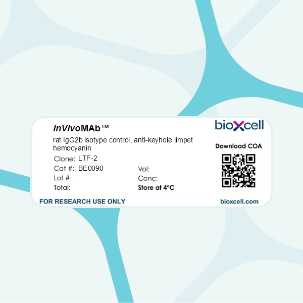
InVivoMAb rat IgG2b isotype control, anti-keyhole limpet hemocyanin
Product Description
Specifications
| Isotype | Rat IgG2b, κ |
|---|---|
| Recommended Dilution Buffer | InVivoPure pH 7.0 Dilution Buffer |
| Conjugation | This product is unconjugated. Conjugation is available via our Antibody Conjugation Services. |
| Formulation |
PBS, pH 7.0 Contains no stabilizers or preservatives |
| Endotoxin |
≤1EU/mg (≤0.001EU/μg) Determined by LAL assay |
| Purity |
≥95% Determined by SDS-PAGE |
| Sterility | 0.2 µm filtration |
| Production | Purified from cell culture supernatant in an animal-free facility |
| Purification | Protein G |
| RRID | AB_1107780 |
| Molecular Weight | 150 kDa |
| Storage | The antibody solution should be stored at the stock concentration at 4°C. Do not freeze. |
| Need a Custom Formulation? | See All Antibody Customization Options |
Application References
Zhang, J., et al (2015). "Micro-RNA-155-mediated control of heme oxygenase 1 (HO-1) is required for restoring adaptively tolerant CD4+ T-cell function in rodents" Eur J Immunol 45(3): 829-842.
PubMed
T cells chronically stimulated by a persistent antigen often become dysfunctional and lose effector functions and proliferative capacity. To identify the importance of micro-RNA-155 (miR-155) in this phenomenon, we analyzed mouse miR-155-deficient CD4(+) T cells in a model where the chronic exposure to a systemic antigen led to T-cell functional unresponsiveness. We found that miR-155 was required for restoring function of T cells after programmed death receptor 1 blockade. Heme oxygenase 1 (HO-1) was identified as a specific target of miR-155 and inhibition of HO-1 activity restored the expansion and tissue migration capacity of miR-155(-/-) CD4(+) T cells. Moreover, miR-155-mediated control of HO-1 expression in CD4(+) T cells was shown to sustain in vivo antigen-specific expansion and IL-2 production. Thus, our data identify HO-1 regulation as a mechanism by which miR-155 promotes T-cell-driven inflammation.
Coers, J., et al (2011). "Compensatory T cell responses in IRG-deficient mice prevent sustained Chlamydia trachomatis infections" PLoS Pathog 7(6): e1001346.
PubMed
The obligate intracellular pathogen Chlamydia trachomatis is the most common cause of bacterial sexually transmitted diseases in the United States. In women C. trachomatis can establish persistent genital infections that lead to pelvic inflammatory disease and sterility. In contrast to natural infections in humans, experimentally induced infections with C. trachomatis in mice are rapidly cleared. The cytokine interferon-gamma (IFNgamma) plays a critical role in the clearance of C. trachomatis infections in mice. Because IFNgamma induces an antimicrobial defense system in mice but not in humans that is composed of a large family of Immunity Related GTPases (IRGs), we questioned whether mice deficient in IRG immunity would develop persistent infections with C. trachomatis as observed in human patients. We found that IRG-deficient Irgm1/m3((-/-)) mice transiently develop high bacterial burden post intrauterine infection, but subsequently clear the infection more efficiently than wildtype mice. We show that the delayed but highly effective clearance of intrauterine C. trachomatis infections in Irgm1/m3((-/-)) mice is dependent on an exacerbated CD4(+) T cell response. These findings indicate that the absence of the predominant murine innate effector mechanism restricting C. trachomatis growth inside epithelial cells results in a compensatory adaptive immune response, which is at least in part driven by CD4(+) T cells and prevents the establishment of a persistent infection in mice.
in vivo PD-L1 blockade
Immunofluorescence
Willimsky, G., et al (2013). "Virus-induced hepatocellular carcinomas cause antigen-specific local tolerance" J Clin Invest 123(3): 1032-1043.
PubMed
T cell surveillance is often effective against virus-associated tumors because of their high immunogenicity. It is not clear why surveillance occasionally fails, particularly against hepatitis B virus- or hepatitis C virus-associated hepatocellular carcinoma (HCC). We established a transgenic murine model of virus-induced HCC by hepatocyte-specific adenovirus-induced activation of the oncogenic SV40 large T antigen (TAg). Adenovirus infection induced cytotoxic T lymphocytes (CTLs) targeted against the virus and TAg, leading to clearance of the infected cells. Despite the presence of functional, antigen-specific T cells, a few virus-infected cells escaped immune clearance and progressed to HCC. These cells expressed TAg at levels similar to HCC isolated from neonatal TAg-tolerant mice, suggesting that CTL clearance does not select for cells with low immunogenicity. Virus-infected mice revealed significantly greater T cell infiltration in early-stage HCC compared with that in late-stage HCC, demonstrating progressive local immune suppression through inefficient T cell infiltration. Programmed cell death protein-1 (PD-1) and its ligand PD-L1 were expressed in all TAg-specific CD8+ T cells and HCC, respectively, which contributed to local tumor-antigen-specific tolerance. Thus, we have developed a model of virus-induced HCC that may allow for a better understanding of human HCC.
in vivo PD-L1 blockade
Flow Cytometry
Rutigliano, J. A., et al (2014). "Highly pathological influenza A virus infection is associated with augmented expression of PD-1 by functionally compromised virus-specific CD8+ T cells" J Virol 88(3): 1636-1651.
PubMed
One question that continues to challenge influenza A research is why some strains of virus are so devastating compared to their more mild counterparts. We approached this question from an immunological perspective, investigating the CD8(+) T cell response in a mouse model system comparing high- and low-pathological influenza virus infections. Our findings reveal that the early (day 0 to 5) viral titer was not the determining factor in the outcome of disease. Instead, increased numbers of antigen-specific CD8(+) T cells and elevated effector function on a per-cell basis were found in the low-pathological infection and correlated with reduced illness and later-time-point (day 6 to 10) viral titer. High-pathological infection was associated with increased PD-1 expression on influenza virus-specific CD8(+) T cells, and blockade of PD-L1 in vivo led to reduced virus titers and increased CD8(+) T cell numbers in high- but not low-pathological infection, though T cell functionality was not restored. These data show that high-pathological acute influenza virus infection is associated with a dysregulated CD8(+) T cell response, which is likely caused by the more highly inflamed airway microenvironment during the early days of infection. Therapeutic approaches specifically aimed at modulating innate airway inflammation may therefore promote efficient CD8(+) T cell activity. We show that during a severe influenza virus infection, one type of immune cell, the CD8 T cell, is less abundant and less functional than in a more mild infection. This dysregulated T cell phenotype correlates with a lower rate of virus clearance in the severe infection and is partially regulated by the expression of a suppressive coreceptor called PD-1. Treatment with an antibody that blocks PD-1 improves T cell functionality and increases virus clearance.
in vivo PD-L1 blockade
Flow Cytometry
Aloulou, M., et al (2016). "Follicular regulatory T cells can be specific for the immunizing antigen and derive from naive T cells" Nat Commun 7: 10579.
PubMed
T follicular regulatory (Tfr) cells are a subset of Foxp3(+) regulatory T (Treg) cells that form in response to immunization or infection, which localize to the germinal centre where they control the magnitude of the response. Despite an increased interest in the role of Tfr cells in humoral immunity, many fundamental aspects of their biology remain unknown, including whether they recognize self- or foreign antigen. Here we show that Tfr cells can be specific for the immunizing antigen, irrespective of whether it is a self- or foreign antigen. We show that, in addition to developing from thymic derived Treg cells, Tfr cells can also arise from Foxp3(-) precursors in a PD-L1-dependent manner, if the adjuvant used is one that supports T-cell plasticity. These findings have important implications for Tfr cell biology and for improving vaccine efficacy by formulating vaccines that modify the Tfr:Tfh cell ratio.
in vivo T cell depletion
Finkin, S., et al (2015). "Ectopic lymphoid structures function as microniches for tumor progenitor cells in hepatocellular carcinoma" Nat Immunol. doi : 10.1038/ni.3290.
PubMed
Ectopic lymphoid-like structures (ELSs) are often observed in cancer, yet their function is obscure. Although ELSs signify good prognosis in certain malignancies, we found that hepatic ELSs indicated poor prognosis for hepatocellular carcinoma (HCC). We studied an HCC mouse model that displayed abundant ELSs and found that they constituted immunopathological microniches wherein malignant hepatocyte progenitor cells appeared and thrived in a complex cellular and cytokine milieu until gaining self-sufficiency. The egress of progenitor cells and tumor formation were associated with the autocrine production of cytokines previously provided by the niche. ELSs developed via cooperation between the innate immune system and adaptive immune system, an event facilitated by activation of the transcription factor NF-kappaB and abolished by depletion of T cells. Such aberrant immunological foci might represent new targets for cancer therapy.
in vivo PD-L1 blockade
Immunofluorescence
Willimsky, G., et al (2013). "Virus-induced hepatocellular carcinomas cause antigen-specific local tolerance" J Clin Invest 123(3): 1032-1043.
PubMed
T cell surveillance is often effective against virus-associated tumors because of their high immunogenicity. It is not clear why surveillance occasionally fails, particularly against hepatitis B virus- or hepatitis C virus-associated hepatocellular carcinoma (HCC). We established a transgenic murine model of virus-induced HCC by hepatocyte-specific adenovirus-induced activation of the oncogenic SV40 large T antigen (TAg). Adenovirus infection induced cytotoxic T lymphocytes (CTLs) targeted against the virus and TAg, leading to clearance of the infected cells. Despite the presence of functional, antigen-specific T cells, a few virus-infected cells escaped immune clearance and progressed to HCC. These cells expressed TAg at levels similar to HCC isolated from neonatal TAg-tolerant mice, suggesting that CTL clearance does not select for cells with low immunogenicity. Virus-infected mice revealed significantly greater T cell infiltration in early-stage HCC compared with that in late-stage HCC, demonstrating progressive local immune suppression through inefficient T cell infiltration. Programmed cell death protein-1 (PD-1) and its ligand PD-L1 were expressed in all TAg-specific CD8+ T cells and HCC, respectively, which contributed to local tumor-antigen-specific tolerance. Thus, we have developed a model of virus-induced HCC that may allow for a better understanding of human HCC.
in vivo PD-L1 blockade
Flow Cytometry
Rutigliano, J. A., et al (2014). "Highly pathological influenza A virus infection is associated with augmented expression of PD-1 by functionally compromised virus-specific CD8+ T cells" J Virol 88(3): 1636-1651.
PubMed
One question that continues to challenge influenza A research is why some strains of virus are so devastating compared to their more mild counterparts. We approached this question from an immunological perspective, investigating the CD8(+) T cell response in a mouse model system comparing high- and low-pathological influenza virus infections. Our findings reveal that the early (day 0 to 5) viral titer was not the determining factor in the outcome of disease. Instead, increased numbers of antigen-specific CD8(+) T cells and elevated effector function on a per-cell basis were found in the low-pathological infection and correlated with reduced illness and later-time-point (day 6 to 10) viral titer. High-pathological infection was associated with increased PD-1 expression on influenza virus-specific CD8(+) T cells, and blockade of PD-L1 in vivo led to reduced virus titers and increased CD8(+) T cell numbers in high- but not low-pathological infection, though T cell functionality was not restored. These data show that high-pathological acute influenza virus infection is associated with a dysregulated CD8(+) T cell response, which is likely caused by the more highly inflamed airway microenvironment during the early days of infection. Therapeutic approaches specifically aimed at modulating innate airway inflammation may therefore promote efficient CD8(+) T cell activity. We show that during a severe influenza virus infection, one type of immune cell, the CD8 T cell, is less abundant and less functional than in a more mild infection. This dysregulated T cell phenotype correlates with a lower rate of virus clearance in the severe infection and is partially regulated by the expression of a suppressive coreceptor called PD-1. Treatment with an antibody that blocks PD-1 improves T cell functionality and increases virus clearance.
in vivo T cell depletion
Finkin, S., et al (2015). "Ectopic lymphoid structures function as microniches for tumor progenitor cells in hepatocellular carcinoma" Nat Immunol. doi : 10.1038/ni.3290.
PubMed
Ectopic lymphoid-like structures (ELSs) are often observed in cancer, yet their function is obscure. Although ELSs signify good prognosis in certain malignancies, we found that hepatic ELSs indicated poor prognosis for hepatocellular carcinoma (HCC). We studied an HCC mouse model that displayed abundant ELSs and found that they constituted immunopathological microniches wherein malignant hepatocyte progenitor cells appeared and thrived in a complex cellular and cytokine milieu until gaining self-sufficiency. The egress of progenitor cells and tumor formation were associated with the autocrine production of cytokines previously provided by the niche. ELSs developed via cooperation between the innate immune system and adaptive immune system, an event facilitated by activation of the transcription factor NF-kappaB and abolished by depletion of T cells. Such aberrant immunological foci might represent new targets for cancer therapy.
in vivo PD-L1 blockade
Flow Cytometry
Aloulou, M., et al (2016). "Follicular regulatory T cells can be specific for the immunizing antigen and derive from naive T cells" Nat Commun 7: 10579.
PubMed
T follicular regulatory (Tfr) cells are a subset of Foxp3(+) regulatory T (Treg) cells that form in response to immunization or infection, which localize to the germinal centre where they control the magnitude of the response. Despite an increased interest in the role of Tfr cells in humoral immunity, many fundamental aspects of their biology remain unknown, including whether they recognize self- or foreign antigen. Here we show that Tfr cells can be specific for the immunizing antigen, irrespective of whether it is a self- or foreign antigen. We show that, in addition to developing from thymic derived Treg cells, Tfr cells can also arise from Foxp3(-) precursors in a PD-L1-dependent manner, if the adjuvant used is one that supports T-cell plasticity. These findings have important implications for Tfr cell biology and for improving vaccine efficacy by formulating vaccines that modify the Tfr:Tfh cell ratio.
in vivo NK cell depletion
Flow Cytometry
in vivo CD8+ T cell depletion
Flow Cytometry
Kearl, T. J., et al (2013). "Programmed death receptor-1/programmed death receptor ligand-1 blockade after transient lymphodepletion to treat myeloma" J Immunol 190(11): 5620-5628.
PubMed
Early phase clinical trials targeting the programmed death receptor-1/ligand-1 (PD-1/PD-L1) pathway to overcome tumor-mediated immunosuppression have reported promising results for a variety of cancers. This pathway appears to play an important role in the failure of immune reactivity to malignant plasma cells in multiple myeloma patients, as the tumor cells express relatively high levels of PD-L1, and T cells show increased PD-1 expression. In the current study, we demonstrate that PD-1/PD-L1 blockade with a PD-L1-specific Ab elicits rejection of a murine myeloma when combined with lymphodepleting irradiation. This particular combined approach by itself has not previously been shown to be efficacious in other tumor models. The antitumor effect of lymphodepletion/anti-PD-L1 therapy was most robust when tumor Ag-experienced T cells were present either through cell transfer or survival after nonmyeloablative irradiation. In vivo depletion of CD4 or CD8 T cells completely eliminated antitumor efficacy of the lymphodepletion/anti-PD-L1 therapy, indicating that both T cell subsets are necessary for tumor rejection. Elimination of myeloma by T cells occurs relatively quickly as tumor cells in the bone marrow were nearly nondetectable by 5 d after the first anti-PD-L1 treatment, suggesting that antimyeloma reactivity is primarily mediated by preactivated T cells, rather than newly generated myeloma-reactive T cells. Anti-PD-L1 plus lymphodepletion failed to improve survival in two solid tumor models, but demonstrated significant efficacy in two hematologic malignancy models. In summary, our results support the clinical testing of lymphodepletion and PD-1/PD-L1 blockade as a novel approach for improving the survival of patients with multiple myeloma.
in vivo macrophage depletion
in vivo CD40 activation
in vivo LAG-3 neutralization
in vivo blocking of ICOS/ICOSL signaling
Flow Cytometry
Bauche, D., et al (2018). "LAG3(+) Regulatory T Cells Restrain Interleukin-23-Producing CX3CR1(+) Gut-Resident Macrophages during Group 3 Innate Lymphoid Cell-Driven Colitis" Immunity 49(2): 342-352 e345.
PubMed
Interleukin-22 (IL-22)-producing group 3 innate lymphoid cells (ILC3) maintains gut homeostasis but can also promote inflammatory bowel disease (IBD). The regulation of ILC3-dependent colitis remains to be elucidated. Here we show that Foxp3(+) regulatory T cells (Treg cells) prevented ILC3-mediated colitis in an IL-10-independent manner. Treg cells inhibited IL-23 and IL-1beta production from intestinal-resident CX3CR1(+) macrophages but not CD103(+) dendritic cells. Moreover, Treg cells restrained ILC3 production of IL-22 through suppression of CX3CR1(+) macrophage production of IL-23 and IL-1beta. This suppression was contact dependent and was mediated by latent activation gene-3 (LAG-3)-an immune checkpoint receptor-expressed on Treg cells. Engagement of LAG-3 on MHC class II drove profound immunosuppression of CX3CR1(+) tissue-resident macrophages. Our study reveals that the health of the intestinal mucosa is maintained by an axis driven by Treg cells communication with resident macrophages that withhold inflammatory stimuli required for ILC3 function.
in vivo regulatory T cell depletion
Flow Cytometry
Park, H. J., et al (2015). "PD-1 upregulated on regulatory T cells during chronic virus infection enhances the suppression of CD8+ T cell immune response via the interaction with PD-L1 expressed on CD8+ T cells" J Immunol 194(12): 5801-5811.
PubMed
Regulatory T (Treg) cells act as terminators of T cell immuniy during acute phase of viral infection; however, their role and suppressive mechanism in chronic viral infection are not completely understood. In this study, we compared the phenotype and function of Treg cells during acute or chronic infection with lymphocytic choriomeningitis virus. Chronic infection, unlike acute infection, led to a large expansion of Treg cells and their upregulation of programmed death-1 (PD-1). Treg cells from chronically infected mice (chronic Treg cells) displayed greater suppressive capacity for inhibiting both CD8(+) and CD4(+) T cell proliferation and subsequent cytokine production than those from naive or acutely infected mice. A contact between Treg and CD8(+) T cells was necessary for the potent suppression of CD8(+) T cell immune response. More importantly, the suppression required cell-specific expression and interaction of PD-1 on chronic Treg cells and PD-1 ligand on CD8(+) T cells. Our study defines PD-1 upregulated on Treg cells and its interaction with PD-1 ligand on effector T cells as one cause for the potent T cell suppression and proposes the role of PD-1 on Treg cells, in addition to that on exhausted T cells, during chronic viral infection.
in vitro TIM-3 blocking
Flow Cytometry
in vivo CD40 activation
in vitro LAG-3 neutralization
Flow Cytometry
in vitro CD48 blocking
in vivo PD-L2 blockade
in vitro PD-L2 blockade
Erickson, J. J., et al (2014). "Programmed death-1 impairs secondary effector lung CD8(+) T cells during respiratory virus reinfection" J Immunol 193(10): 5108-5117.
PubMed
Reinfections with respiratory viruses are common and cause significant clinical illness, yet precise mechanisms governing this susceptibility are ill defined. Lung Ag-specific CD8(+) T cells (T(CD8)) are impaired during acute viral lower respiratory infection by the inhibitory receptor programmed death-1 (PD-1). To determine whether PD-1 contributes to recurrent infection, we first established a model of reinfection by challenging B cell-deficient mice with human metapneumovirus (HMPV) several weeks after primary infection, and found that HMPV replicated to high titers in the lungs. A robust secondary effector lung TCD8 response was generated during reinfection, but these cells were more impaired and more highly expressed the inhibitory receptors PD-1, LAG-3, and 2B4 than primary T(CD8). In vitro blockade demonstrated that PD-1 was the dominant inhibitory receptor early after reinfection. In vivo therapeutic PD-1 blockade during HMPV reinfection restored lung T(CD8) effector functions (i.e., degranulation and cytokine production) and enhanced viral clearance. PD-1 also limited the protective efficacy of HMPV epitope-specific peptide vaccination and impaired lung T(CD8) during heterotypic influenza virus challenge infection. Our results indicate that PD-1 signaling may contribute to respiratory virus reinfection and evasion of vaccine-elicited immune responses. These results have important implications for the design of effective vaccines against respiratory viruses.
in vivo CD8+ T cell depletion
in vitro T cell stimulation/activation
Sledzinska, A., et al (2013). "TGF-beta signalling is required for CD4(+) T cell homeostasis but dispensable for regulatory T cell function" PLoS Biol 11(10): e1001674.
PubMed
TGF-beta is widely held to be critical for the maintenance and function of regulatory T (T(reg)) cells and thus peripheral tolerance. This is highlighted by constitutive ablation of TGF-beta receptor (TR) during thymic development in mice, which leads to a lethal autoimmune syndrome. Here we describe that TGF-beta-driven peripheral tolerance is not regulated by TGF-beta signalling on mature CD4(+) T cells. Inducible TR2 ablation specifically on CD4(+) T cells did not result in a lethal autoinflammation. Transfer of these TR2-deficient CD4(+) T cells to lymphopenic recipients resulted in colitis, but not overt autoimmunity. In contrast, thymic ablation of TR2 in combination with lymphopenia led to lethal multi-organ inflammation. Interestingly, deletion of TR2 on mature CD4(+) T cells does not result in the collapse of the T(reg) cell population as observed in constitutive models. Instead, a pronounced enlargement of both regulatory and effector memory T cell pools was observed. This expansion is cell-intrinsic and seems to be caused by increased T cell receptor sensitivity independently of common gamma chain-dependent cytokine signals. The expression of Foxp3 and other regulatory T cells markers was not dependent on TGF-beta signalling and the TR2-deficient T(reg) cells retained their suppressive function both in vitro and in vivo. In summary, absence of TGF-beta signalling on mature CD4(+) T cells is not responsible for breakdown of peripheral tolerance, but rather controls homeostasis of mature T cells in adult mice.
in vitro TIM-3 blocking
Flow Cytometry
in vivo CD40 activation
in vitro LAG-3 neutralization
Flow Cytometry
in vitro CD48 blocking
in vivo PD-L2 blockade
in vitro PD-L2 blockade
Erickson, J. J., et al (2014). "Programmed death-1 impairs secondary effector lung CD8(+) T cells during respiratory virus reinfection" J Immunol 193(10): 5108-5117.
PubMed
Reinfections with respiratory viruses are common and cause significant clinical illness, yet precise mechanisms governing this susceptibility are ill defined. Lung Ag-specific CD8(+) T cells (T(CD8)) are impaired during acute viral lower respiratory infection by the inhibitory receptor programmed death-1 (PD-1). To determine whether PD-1 contributes to recurrent infection, we first established a model of reinfection by challenging B cell-deficient mice with human metapneumovirus (HMPV) several weeks after primary infection, and found that HMPV replicated to high titers in the lungs. A robust secondary effector lung TCD8 response was generated during reinfection, but these cells were more impaired and more highly expressed the inhibitory receptors PD-1, LAG-3, and 2B4 than primary T(CD8). In vitro blockade demonstrated that PD-1 was the dominant inhibitory receptor early after reinfection. In vivo therapeutic PD-1 blockade during HMPV reinfection restored lung T(CD8) effector functions (i.e., degranulation and cytokine production) and enhanced viral clearance. PD-1 also limited the protective efficacy of HMPV epitope-specific peptide vaccination and impaired lung T(CD8) during heterotypic influenza virus challenge infection. Our results indicate that PD-1 signaling may contribute to respiratory virus reinfection and evasion of vaccine-elicited immune responses. These results have important implications for the design of effective vaccines against respiratory viruses.
in vivo depletion of Gr-1+ myeloid cells
Flow Cytometry
van der Merwe, M., et al (2013). "Recipient myeloid-derived immunomodulatory cells induce PD-1 ligand-dependent donor CD4+Foxp3+ regulatory T cell proliferation and donor-recipient immune tolerance after murine nonmyeloablative bone marrow transplant
PubMed
We showed previously that nonmyeloablative total lymphoid irradiation/rabbit anti-thymocyte serum (TLI/ATS) conditioning facilitates potent donor-recipient immune tolerance following bone marrow transplantation (BMT) across MHC barriers via recipient invariant NKT (iNKT) cell-derived IL-4-dependent expansion of donor Foxp3(+) naturally occurring regulatory T cells (nTregs). In this study, we report a more specific mechanism. Wild-type (WT) BALB/c (H-2(d)) hosts were administered TLI/ATS and BMT from WT or STAT6(-/-) C57BL/6 (H-2(b)) donors. Following STAT6(-/-) BMT, donor nTregs demonstrated no loss of proliferation in vivo, indicating that an IL-4-responsive population in the recipient, rather than the donor, drives donor nTreg proliferation. In graft-versus-host disease (GVHD) target organs, three recipient CD11b(+) cell subsets (Gr-1(high)CD11c(-), Gr-1(int)CD11c(-), and Gr-1(low)CD11c(+)) were enriched early after TLI/ATS + BMT versus total body irradiation/ATS + BMT. Gr-1(low)CD11c(+) cells induced potent H-2K(b+)CD4(+)Foxp3(+) nTreg proliferation in vitro in 72-h MLRs. Gr-1(low)CD11c(+) cells were reduced significantly in STAT6(-/-) and iNKT cell-deficient Jalpha18(-/-) BALB/c recipients after TLI/ATS + BMT. Depletion of CD11b(+) cells resulted in severe acute GVHD, and adoptive transfer of WT Gr-1(low)CD11c(+) cells to Jalpha18(-/-) BALB/c recipients of TLI/ATS + BMT restored day-6 donor Foxp3(+) nTreg proliferation and protection from CD8 effector T cell-mediated GVHD. Blockade of programmed death ligand 1 and 2, but not CD40, TGF-beta signaling, arginase 1, or iNOS, inhibited nTreg proliferation in cocultures of recipient-derived Gr-1(low)CD11c(+) cells with donor nTregs. Through iNKT-dependent Th2 polarization, myeloid-derived immunomodulatory dendritic cells are expanded after nonmyeloablative TLI/ATS conditioning and allogeneic BMT, induce PD-1 ligand-dependent donor nTreg proliferation, and maintain potent graft-versus-host immune tolerance.
in vivo blocking of PD-1/PD-L signaling
in vivo PD-L1 blockade
in vivo CTLA-4 neutralization
in vivo CD8+ T cell depletion
Twyman-Saint Victor, C., et al (2015). "Radiation and dual checkpoint blockade activate non-redundant immune mechanisms in cancer" Nature 520(7547): 373-377.
PubMed
Immune checkpoint inhibitors result in impressive clinical responses, but optimal results will require combination with each other and other therapies. This raises fundamental questions about mechanisms of non-redundancy and resistance. Here we report major tumour regressions in a subset of patients with metastatic melanoma treated with an anti-CTLA4 antibody (anti-CTLA4) and radiation, and reproduced this effect in mouse models. Although combined treatment improved responses in irradiated and unirradiated tumours, resistance was common. Unbiased analyses of mice revealed that resistance was due to upregulation of PD-L1 on melanoma cells and associated with T-cell exhaustion. Accordingly, optimal response in melanoma and other cancer types requires radiation, anti-CTLA4 and anti-PD-L1/PD-1. Anti-CTLA4 predominantly inhibits T-regulatory cells (Treg cells), thereby increasing the CD8 T-cell to Treg (CD8/Treg) ratio. Radiation enhances the diversity of the T-cell receptor (TCR) repertoire of intratumoral T cells. Together, anti-CTLA4 promotes expansion of T cells, while radiation shapes the TCR repertoire of the expanded peripheral clones. Addition of PD-L1 blockade reverses T-cell exhaustion to mitigate depression in the CD8/Treg ratio and further encourages oligoclonal T-cell expansion. Similarly to results from mice, patients on our clinical trial with melanoma showing high PD-L1 did not respond to radiation plus anti-CTLA4, demonstrated persistent T-cell exhaustion, and rapidly progressed. Thus, PD-L1 on melanoma cells allows tumours to escape anti-CTLA4-based therapy, and the combination of radiation, anti-CTLA4 and anti-PD-L1 promotes response and immunity through distinct mechanisms.
in vivo CD8+ T cell depletion
in vitro T cell stimulation/activation
Sledzinska, A., et al (2013). "TGF-beta signalling is required for CD4(+) T cell homeostasis but dispensable for regulatory T cell function" PLoS Biol 11(10): e1001674.
PubMed
TGF-beta is widely held to be critical for the maintenance and function of regulatory T (T(reg)) cells and thus peripheral tolerance. This is highlighted by constitutive ablation of TGF-beta receptor (TR) during thymic development in mice, which leads to a lethal autoimmune syndrome. Here we describe that TGF-beta-driven peripheral tolerance is not regulated by TGF-beta signalling on mature CD4(+) T cells. Inducible TR2 ablation specifically on CD4(+) T cells did not result in a lethal autoinflammation. Transfer of these TR2-deficient CD4(+) T cells to lymphopenic recipients resulted in colitis, but not overt autoimmunity. In contrast, thymic ablation of TR2 in combination with lymphopenia led to lethal multi-organ inflammation. Interestingly, deletion of TR2 on mature CD4(+) T cells does not result in the collapse of the T(reg) cell population as observed in constitutive models. Instead, a pronounced enlargement of both regulatory and effector memory T cell pools was observed. This expansion is cell-intrinsic and seems to be caused by increased T cell receptor sensitivity independently of common gamma chain-dependent cytokine signals. The expression of Foxp3 and other regulatory T cells markers was not dependent on TGF-beta signalling and the TR2-deficient T(reg) cells retained their suppressive function both in vitro and in vivo. In summary, absence of TGF-beta signalling on mature CD4(+) T cells is not responsible for breakdown of peripheral tolerance, but rather controls homeostasis of mature T cells in adult mice.
in vivo blocking of PD-1/PD-L signaling
in vivo PD-L1 blockade
in vivo CTLA-4 neutralization
in vivo CD8+ T cell depletion
Twyman-Saint Victor, C., et al (2015). "Radiation and dual checkpoint blockade activate non-redundant immune mechanisms in cancer" Nature 520(7547): 373-377.
PubMed
Immune checkpoint inhibitors result in impressive clinical responses, but optimal results will require combination with each other and other therapies. This raises fundamental questions about mechanisms of non-redundancy and resistance. Here we report major tumour regressions in a subset of patients with metastatic melanoma treated with an anti-CTLA4 antibody (anti-CTLA4) and radiation, and reproduced this effect in mouse models. Although combined treatment improved responses in irradiated and unirradiated tumours, resistance was common. Unbiased analyses of mice revealed that resistance was due to upregulation of PD-L1 on melanoma cells and associated with T-cell exhaustion. Accordingly, optimal response in melanoma and other cancer types requires radiation, anti-CTLA4 and anti-PD-L1/PD-1. Anti-CTLA4 predominantly inhibits T-regulatory cells (Treg cells), thereby increasing the CD8 T-cell to Treg (CD8/Treg) ratio. Radiation enhances the diversity of the T-cell receptor (TCR) repertoire of intratumoral T cells. Together, anti-CTLA4 promotes expansion of T cells, while radiation shapes the TCR repertoire of the expanded peripheral clones. Addition of PD-L1 blockade reverses T-cell exhaustion to mitigate depression in the CD8/Treg ratio and further encourages oligoclonal T-cell expansion. Similarly to results from mice, patients on our clinical trial with melanoma showing high PD-L1 did not respond to radiation plus anti-CTLA4, demonstrated persistent T-cell exhaustion, and rapidly progressed. Thus, PD-L1 on melanoma cells allows tumours to escape anti-CTLA4-based therapy, and the combination of radiation, anti-CTLA4 and anti-PD-L1 promotes response and immunity through distinct mechanisms.
in vivo depletion of Gr-1+ myeloid cells
Flow Cytometry
van der Merwe, M., et al (2013). "Recipient myeloid-derived immunomodulatory cells induce PD-1 ligand-dependent donor CD4+Foxp3+ regulatory T cell proliferation and donor-recipient immune tolerance after murine nonmyeloablative bone marrow transplant
PubMed
We showed previously that nonmyeloablative total lymphoid irradiation/rabbit anti-thymocyte serum (TLI/ATS) conditioning facilitates potent donor-recipient immune tolerance following bone marrow transplantation (BMT) across MHC barriers via recipient invariant NKT (iNKT) cell-derived IL-4-dependent expansion of donor Foxp3(+) naturally occurring regulatory T cells (nTregs). In this study, we report a more specific mechanism. Wild-type (WT) BALB/c (H-2(d)) hosts were administered TLI/ATS and BMT from WT or STAT6(-/-) C57BL/6 (H-2(b)) donors. Following STAT6(-/-) BMT, donor nTregs demonstrated no loss of proliferation in vivo, indicating that an IL-4-responsive population in the recipient, rather than the donor, drives donor nTreg proliferation. In graft-versus-host disease (GVHD) target organs, three recipient CD11b(+) cell subsets (Gr-1(high)CD11c(-), Gr-1(int)CD11c(-), and Gr-1(low)CD11c(+)) were enriched early after TLI/ATS + BMT versus total body irradiation/ATS + BMT. Gr-1(low)CD11c(+) cells induced potent H-2K(b+)CD4(+)Foxp3(+) nTreg proliferation in vitro in 72-h MLRs. Gr-1(low)CD11c(+) cells were reduced significantly in STAT6(-/-) and iNKT cell-deficient Jalpha18(-/-) BALB/c recipients after TLI/ATS + BMT. Depletion of CD11b(+) cells resulted in severe acute GVHD, and adoptive transfer of WT Gr-1(low)CD11c(+) cells to Jalpha18(-/-) BALB/c recipients of TLI/ATS + BMT restored day-6 donor Foxp3(+) nTreg proliferation and protection from CD8 effector T cell-mediated GVHD. Blockade of programmed death ligand 1 and 2, but not CD40, TGF-beta signaling, arginase 1, or iNOS, inhibited nTreg proliferation in cocultures of recipient-derived Gr-1(low)CD11c(+) cells with donor nTregs. Through iNKT-dependent Th2 polarization, myeloid-derived immunomodulatory dendritic cells are expanded after nonmyeloablative TLI/ATS conditioning and allogeneic BMT, induce PD-1 ligand-dependent donor nTreg proliferation, and maintain potent graft-versus-host immune tolerance.
in vivo CTLA-4 neutralization
in vivo CD8+ T cell depletion
in vivo blocking of PD-1/PD-L signaling
Triplett, T. A., et al (2018). "Reversal of indoleamine 2,3-dioxygenase-mediated cancer immune suppression by systemic kynurenine depletion with a therapeutic enzyme" Nat Biotechnol 36(8): 758-764.
PubMed
Increased tryptophan (Trp) catabolism in the tumor microenvironment (TME) can mediate immune suppression by upregulation of interferon (IFN)-gamma-inducible indoleamine 2,3-dioxygenase (IDO1) and/or ectopic expression of the predominantly liver-restricted enzyme tryptophan 2,3-dioxygenase (TDO). Whether these effects are due to Trp depletion in the TME or mediated by the accumulation of the IDO1 and/or TDO (hereafter referred to as IDO1/TDO) product kynurenine (Kyn) remains controversial. Here we show that administration of a pharmacologically optimized enzyme (PEGylated kynureninase; hereafter referred to as PEG-KYNase) that degrades Kyn into immunologically inert, nontoxic and readily cleared metabolites inhibits tumor growth. Enzyme treatment was associated with a marked increase in the tumor infiltration and proliferation of polyfunctional CD8(+) lymphocytes. We show that PEG-KYNase administration had substantial therapeutic effects when combined with approved checkpoint inhibitors or with a cancer vaccine for the treatment of large B16-F10 melanoma, 4T1 breast carcinoma or CT26 colon carcinoma tumors. PEG-KYNase mediated prolonged depletion of Kyn in the TME and reversed the modulatory effects of IDO1/TDO upregulation in the TME.
in vivo CD4+ T cell depletion
Steel, C. D., et al (2014). "Role of peripheral immune response in microglia activation and regulation of brain chemokine and proinflammatory cytokine responses induced during VSV encephalitis" J Neuroimmunol 267(1-2): 50-60.
PubMed
We report herein that neuroinvasion by vesicular stomatitis virus (VSV) activates microglia and induces a peripheral dendritic cell (DC)-dependent inflammatory response in the central nervous system (CNS). VSV neuroinvasion rapidly induces multiple brain chemokine and proinflammatory cytokine mRNAs that display bimodal kinetics. Peripheral DC ablation or T cell depletion suppresses the second wave of this response demonstrating that infiltrating T cells are primarily responsible for the bimodal characteristics of this response. The robust infiltrate associated with VSV encephalitis likely depends on sustained production of brain CCL19 and CCR7 expression on infiltrating inflammatory cells.
Bauche, D., et al (2018). "LAG3(+) Regulatory T Cells Restrain Interleukin-23-Producing CX3CR1(+) Gut-Resident Macrophages during Group 3 Innate Lymphoid Cell-Driven Colitis" Immunity 49(2): 342-352 e345.
PubMed
Interleukin-22 (IL-22)-producing group 3 innate lymphoid cells (ILC3) maintains gut homeostasis but can also promote inflammatory bowel disease (IBD). The regulation of ILC3-dependent colitis remains to be elucidated. Here we show that Foxp3(+) regulatory T cells (Treg cells) prevented ILC3-mediated colitis in an IL-10-independent manner. Treg cells inhibited IL-23 and IL-1beta production from intestinal-resident CX3CR1(+) macrophages but not CD103(+) dendritic cells. Moreover, Treg cells restrained ILC3 production of IL-22 through suppression of CX3CR1(+) macrophage production of IL-23 and IL-1beta. This suppression was contact dependent and was mediated by latent activation gene-3 (LAG-3)-an immune checkpoint receptor-expressed on Treg cells. Engagement of LAG-3 on MHC class II drove profound immunosuppression of CX3CR1(+) tissue-resident macrophages. Our study reveals that the health of the intestinal mucosa is maintained by an axis driven by Treg cells communication with resident macrophages that withhold inflammatory stimuli required for ILC3 function.
Triplett, T. A., et al (2018). "Reversal of indoleamine 2,3-dioxygenase-mediated cancer immune suppression by systemic kynurenine depletion with a therapeutic enzyme" Nat Biotechnol 36(8): 758-764.
PubMed
Increased tryptophan (Trp) catabolism in the tumor microenvironment (TME) can mediate immune suppression by upregulation of interferon (IFN)-gamma-inducible indoleamine 2,3-dioxygenase (IDO1) and/or ectopic expression of the predominantly liver-restricted enzyme tryptophan 2,3-dioxygenase (TDO). Whether these effects are due to Trp depletion in the TME or mediated by the accumulation of the IDO1 and/or TDO (hereafter referred to as IDO1/TDO) product kynurenine (Kyn) remains controversial. Here we show that administration of a pharmacologically optimized enzyme (PEGylated kynureninase; hereafter referred to as PEG-KYNase) that degrades Kyn into immunologically inert, nontoxic and readily cleared metabolites inhibits tumor growth. Enzyme treatment was associated with a marked increase in the tumor infiltration and proliferation of polyfunctional CD8(+) lymphocytes. We show that PEG-KYNase administration had substantial therapeutic effects when combined with approved checkpoint inhibitors or with a cancer vaccine for the treatment of large B16-F10 melanoma, 4T1 breast carcinoma or CT26 colon carcinoma tumors. PEG-KYNase mediated prolonged depletion of Kyn in the TME and reversed the modulatory effects of IDO1/TDO upregulation in the TME.
Aloulou, M., et al (2016). "Follicular regulatory T cells can be specific for the immunizing antigen and derive from naive T cells" Nat Commun 7: 10579.
PubMed
T follicular regulatory (Tfr) cells are a subset of Foxp3(+) regulatory T (Treg) cells that form in response to immunization or infection, which localize to the germinal centre where they control the magnitude of the response. Despite an increased interest in the role of Tfr cells in humoral immunity, many fundamental aspects of their biology remain unknown, including whether they recognize self- or foreign antigen. Here we show that Tfr cells can be specific for the immunizing antigen, irrespective of whether it is a self- or foreign antigen. We show that, in addition to developing from thymic derived Treg cells, Tfr cells can also arise from Foxp3(-) precursors in a PD-L1-dependent manner, if the adjuvant used is one that supports T-cell plasticity. These findings have important implications for Tfr cell biology and for improving vaccine efficacy by formulating vaccines that modify the Tfr:Tfh cell ratio.
Twyman-Saint Victor, C., et al (2015). "Radiation and dual checkpoint blockade activate non-redundant immune mechanisms in cancer" Nature 520(7547): 373-377.
PubMed
Immune checkpoint inhibitors result in impressive clinical responses, but optimal results will require combination with each other and other therapies. This raises fundamental questions about mechanisms of non-redundancy and resistance. Here we report major tumour regressions in a subset of patients with metastatic melanoma treated with an anti-CTLA4 antibody (anti-CTLA4) and radiation, and reproduced this effect in mouse models. Although combined treatment improved responses in irradiated and unirradiated tumours, resistance was common. Unbiased analyses of mice revealed that resistance was due to upregulation of PD-L1 on melanoma cells and associated with T-cell exhaustion. Accordingly, optimal response in melanoma and other cancer types requires radiation, anti-CTLA4 and anti-PD-L1/PD-1. Anti-CTLA4 predominantly inhibits T-regulatory cells (Treg cells), thereby increasing the CD8 T-cell to Treg (CD8/Treg) ratio. Radiation enhances the diversity of the T-cell receptor (TCR) repertoire of intratumoral T cells. Together, anti-CTLA4 promotes expansion of T cells, while radiation shapes the TCR repertoire of the expanded peripheral clones. Addition of PD-L1 blockade reverses T-cell exhaustion to mitigate depression in the CD8/Treg ratio and further encourages oligoclonal T-cell expansion. Similarly to results from mice, patients on our clinical trial with melanoma showing high PD-L1 did not respond to radiation plus anti-CTLA4, demonstrated persistent T-cell exhaustion, and rapidly progressed. Thus, PD-L1 on melanoma cells allows tumours to escape anti-CTLA4-based therapy, and the combination of radiation, anti-CTLA4 and anti-PD-L1 promotes response and immunity through distinct mechanisms.
Zhang, J., et al (2015). "Micro-RNA-155-mediated control of heme oxygenase 1 (HO-1) is required for restoring adaptively tolerant CD4+ T-cell function in rodents" Eur J Immunol 45(3): 829-842.
PubMed
T cells chronically stimulated by a persistent antigen often become dysfunctional and lose effector functions and proliferative capacity. To identify the importance of micro-RNA-155 (miR-155) in this phenomenon, we analyzed mouse miR-155-deficient CD4(+) T cells in a model where the chronic exposure to a systemic antigen led to T-cell functional unresponsiveness. We found that miR-155 was required for restoring function of T cells after programmed death receptor 1 blockade. Heme oxygenase 1 (HO-1) was identified as a specific target of miR-155 and inhibition of HO-1 activity restored the expansion and tissue migration capacity of miR-155(-/-) CD4(+) T cells. Moreover, miR-155-mediated control of HO-1 expression in CD4(+) T cells was shown to sustain in vivo antigen-specific expansion and IL-2 production. Thus, our data identify HO-1 regulation as a mechanism by which miR-155 promotes T-cell-driven inflammation.
Park, H. J., et al (2015). "PD-1 upregulated on regulatory T cells during chronic virus infection enhances the suppression of CD8+ T cell immune response via the interaction with PD-L1 expressed on CD8+ T cells" J Immunol 194(12): 5801-5811.
PubMed
Regulatory T (Treg) cells act as terminators of T cell immuniy during acute phase of viral infection; however, their role and suppressive mechanism in chronic viral infection are not completely understood. In this study, we compared the phenotype and function of Treg cells during acute or chronic infection with lymphocytic choriomeningitis virus. Chronic infection, unlike acute infection, led to a large expansion of Treg cells and their upregulation of programmed death-1 (PD-1). Treg cells from chronically infected mice (chronic Treg cells) displayed greater suppressive capacity for inhibiting both CD8(+) and CD4(+) T cell proliferation and subsequent cytokine production than those from naive or acutely infected mice. A contact between Treg and CD8(+) T cells was necessary for the potent suppression of CD8(+) T cell immune response. More importantly, the suppression required cell-specific expression and interaction of PD-1 on chronic Treg cells and PD-1 ligand on CD8(+) T cells. Our study defines PD-1 upregulated on Treg cells and its interaction with PD-1 ligand on effector T cells as one cause for the potent T cell suppression and proposes the role of PD-1 on Treg cells, in addition to that on exhausted T cells, during chronic viral infection.
Finkin, S., et al (2015). "Ectopic lymphoid structures function as microniches for tumor progenitor cells in hepatocellular carcinoma" Nat Immunol. doi : 10.1038/ni.3290.
PubMed
Ectopic lymphoid-like structures (ELSs) are often observed in cancer, yet their function is obscure. Although ELSs signify good prognosis in certain malignancies, we found that hepatic ELSs indicated poor prognosis for hepatocellular carcinoma (HCC). We studied an HCC mouse model that displayed abundant ELSs and found that they constituted immunopathological microniches wherein malignant hepatocyte progenitor cells appeared and thrived in a complex cellular and cytokine milieu until gaining self-sufficiency. The egress of progenitor cells and tumor formation were associated with the autocrine production of cytokines previously provided by the niche. ELSs developed via cooperation between the innate immune system and adaptive immune system, an event facilitated by activation of the transcription factor NF-kappaB and abolished by depletion of T cells. Such aberrant immunological foci might represent new targets for cancer therapy.
Rutigliano, J. A., et al (2014). "Highly pathological influenza A virus infection is associated with augmented expression of PD-1 by functionally compromised virus-specific CD8+ T cells" J Virol 88(3): 1636-1651.
PubMed
One question that continues to challenge influenza A research is why some strains of virus are so devastating compared to their more mild counterparts. We approached this question from an immunological perspective, investigating the CD8(+) T cell response in a mouse model system comparing high- and low-pathological influenza virus infections. Our findings reveal that the early (day 0 to 5) viral titer was not the determining factor in the outcome of disease. Instead, increased numbers of antigen-specific CD8(+) T cells and elevated effector function on a per-cell basis were found in the low-pathological infection and correlated with reduced illness and later-time-point (day 6 to 10) viral titer. High-pathological infection was associated with increased PD-1 expression on influenza virus-specific CD8(+) T cells, and blockade of PD-L1 in vivo led to reduced virus titers and increased CD8(+) T cell numbers in high- but not low-pathological infection, though T cell functionality was not restored. These data show that high-pathological acute influenza virus infection is associated with a dysregulated CD8(+) T cell response, which is likely caused by the more highly inflamed airway microenvironment during the early days of infection. Therapeutic approaches specifically aimed at modulating innate airway inflammation may therefore promote efficient CD8(+) T cell activity. We show that during a severe influenza virus infection, one type of immune cell, the CD8 T cell, is less abundant and less functional than in a more mild infection. This dysregulated T cell phenotype correlates with a lower rate of virus clearance in the severe infection and is partially regulated by the expression of a suppressive coreceptor called PD-1. Treatment with an antibody that blocks PD-1 improves T cell functionality and increases virus clearance.
Erickson, J. J., et al (2014). "Programmed death-1 impairs secondary effector lung CD8(+) T cells during respiratory virus reinfection" J Immunol 193(10): 5108-5117.
PubMed
Reinfections with respiratory viruses are common and cause significant clinical illness, yet precise mechanisms governing this susceptibility are ill defined. Lung Ag-specific CD8(+) T cells (T(CD8)) are impaired during acute viral lower respiratory infection by the inhibitory receptor programmed death-1 (PD-1). To determine whether PD-1 contributes to recurrent infection, we first established a model of reinfection by challenging B cell-deficient mice with human metapneumovirus (HMPV) several weeks after primary infection, and found that HMPV replicated to high titers in the lungs. A robust secondary effector lung TCD8 response was generated during reinfection, but these cells were more impaired and more highly expressed the inhibitory receptors PD-1, LAG-3, and 2B4 than primary T(CD8). In vitro blockade demonstrated that PD-1 was the dominant inhibitory receptor early after reinfection. In vivo therapeutic PD-1 blockade during HMPV reinfection restored lung T(CD8) effector functions (i.e., degranulation and cytokine production) and enhanced viral clearance. PD-1 also limited the protective efficacy of HMPV epitope-specific peptide vaccination and impaired lung T(CD8) during heterotypic influenza virus challenge infection. Our results indicate that PD-1 signaling may contribute to respiratory virus reinfection and evasion of vaccine-elicited immune responses. These results have important implications for the design of effective vaccines against respiratory viruses.
van der Merwe, M., et al (2013). "Recipient myeloid-derived immunomodulatory cells induce PD-1 ligand-dependent donor CD4+Foxp3+ regulatory T cell proliferation and donor-recipient immune tolerance after murine nonmyeloablative bone marrow transplant
PubMed
We showed previously that nonmyeloablative total lymphoid irradiation/rabbit anti-thymocyte serum (TLI/ATS) conditioning facilitates potent donor-recipient immune tolerance following bone marrow transplantation (BMT) across MHC barriers via recipient invariant NKT (iNKT) cell-derived IL-4-dependent expansion of donor Foxp3(+) naturally occurring regulatory T cells (nTregs). In this study, we report a more specific mechanism. Wild-type (WT) BALB/c (H-2(d)) hosts were administered TLI/ATS and BMT from WT or STAT6(-/-) C57BL/6 (H-2(b)) donors. Following STAT6(-/-) BMT, donor nTregs demonstrated no loss of proliferation in vivo, indicating that an IL-4-responsive population in the recipient, rather than the donor, drives donor nTreg proliferation. In graft-versus-host disease (GVHD) target organs, three recipient CD11b(+) cell subsets (Gr-1(high)CD11c(-), Gr-1(int)CD11c(-), and Gr-1(low)CD11c(+)) were enriched early after TLI/ATS + BMT versus total body irradiation/ATS + BMT. Gr-1(low)CD11c(+) cells induced potent H-2K(b+)CD4(+)Foxp3(+) nTreg proliferation in vitro in 72-h MLRs. Gr-1(low)CD11c(+) cells were reduced significantly in STAT6(-/-) and iNKT cell-deficient Jalpha18(-/-) BALB/c recipients after TLI/ATS + BMT. Depletion of CD11b(+) cells resulted in severe acute GVHD, and adoptive transfer of WT Gr-1(low)CD11c(+) cells to Jalpha18(-/-) BALB/c recipients of TLI/ATS + BMT restored day-6 donor Foxp3(+) nTreg proliferation and protection from CD8 effector T cell-mediated GVHD. Blockade of programmed death ligand 1 and 2, but not CD40, TGF-beta signaling, arginase 1, or iNOS, inhibited nTreg proliferation in cocultures of recipient-derived Gr-1(low)CD11c(+) cells with donor nTregs. Through iNKT-dependent Th2 polarization, myeloid-derived immunomodulatory dendritic cells are expanded after nonmyeloablative TLI/ATS conditioning and allogeneic BMT, induce PD-1 ligand-dependent donor nTreg proliferation, and maintain potent graft-versus-host immune tolerance.
Willimsky, G., et al (2013). "Virus-induced hepatocellular carcinomas cause antigen-specific local tolerance" J Clin Invest 123(3): 1032-1043.
PubMed
T cell surveillance is often effective against virus-associated tumors because of their high immunogenicity. It is not clear why surveillance occasionally fails, particularly against hepatitis B virus- or hepatitis C virus-associated hepatocellular carcinoma (HCC). We established a transgenic murine model of virus-induced HCC by hepatocyte-specific adenovirus-induced activation of the oncogenic SV40 large T antigen (TAg). Adenovirus infection induced cytotoxic T lymphocytes (CTLs) targeted against the virus and TAg, leading to clearance of the infected cells. Despite the presence of functional, antigen-specific T cells, a few virus-infected cells escaped immune clearance and progressed to HCC. These cells expressed TAg at levels similar to HCC isolated from neonatal TAg-tolerant mice, suggesting that CTL clearance does not select for cells with low immunogenicity. Virus-infected mice revealed significantly greater T cell infiltration in early-stage HCC compared with that in late-stage HCC, demonstrating progressive local immune suppression through inefficient T cell infiltration. Programmed cell death protein-1 (PD-1) and its ligand PD-L1 were expressed in all TAg-specific CD8+ T cells and HCC, respectively, which contributed to local tumor-antigen-specific tolerance. Thus, we have developed a model of virus-induced HCC that may allow for a better understanding of human HCC.
Kearl, T. J., et al (2013). "Programmed death receptor-1/programmed death receptor ligand-1 blockade after transient lymphodepletion to treat myeloma" J Immunol 190(11): 5620-5628.
PubMed
Early phase clinical trials targeting the programmed death receptor-1/ligand-1 (PD-1/PD-L1) pathway to overcome tumor-mediated immunosuppression have reported promising results for a variety of cancers. This pathway appears to play an important role in the failure of immune reactivity to malignant plasma cells in multiple myeloma patients, as the tumor cells express relatively high levels of PD-L1, and T cells show increased PD-1 expression. In the current study, we demonstrate that PD-1/PD-L1 blockade with a PD-L1-specific Ab elicits rejection of a murine myeloma when combined with lymphodepleting irradiation. This particular combined approach by itself has not previously been shown to be efficacious in other tumor models. The antitumor effect of lymphodepletion/anti-PD-L1 therapy was most robust when tumor Ag-experienced T cells were present either through cell transfer or survival after nonmyeloablative irradiation. In vivo depletion of CD4 or CD8 T cells completely eliminated antitumor efficacy of the lymphodepletion/anti-PD-L1 therapy, indicating that both T cell subsets are necessary for tumor rejection. Elimination of myeloma by T cells occurs relatively quickly as tumor cells in the bone marrow were nearly nondetectable by 5 d after the first anti-PD-L1 treatment, suggesting that antimyeloma reactivity is primarily mediated by preactivated T cells, rather than newly generated myeloma-reactive T cells. Anti-PD-L1 plus lymphodepletion failed to improve survival in two solid tumor models, but demonstrated significant efficacy in two hematologic malignancy models. In summary, our results support the clinical testing of lymphodepletion and PD-1/PD-L1 blockade as a novel approach for improving the survival of patients with multiple myeloma.
Sledzinska, A., et al (2013). "TGF-beta signalling is required for CD4(+) T cell homeostasis but dispensable for regulatory T cell function" PLoS Biol 11(10): e1001674.
PubMed
TGF-beta is widely held to be critical for the maintenance and function of regulatory T (T(reg)) cells and thus peripheral tolerance. This is highlighted by constitutive ablation of TGF-beta receptor (TR) during thymic development in mice, which leads to a lethal autoimmune syndrome. Here we describe that TGF-beta-driven peripheral tolerance is not regulated by TGF-beta signalling on mature CD4(+) T cells. Inducible TR2 ablation specifically on CD4(+) T cells did not result in a lethal autoinflammation. Transfer of these TR2-deficient CD4(+) T cells to lymphopenic recipients resulted in colitis, but not overt autoimmunity. In contrast, thymic ablation of TR2 in combination with lymphopenia led to lethal multi-organ inflammation. Interestingly, deletion of TR2 on mature CD4(+) T cells does not result in the collapse of the T(reg) cell population as observed in constitutive models. Instead, a pronounced enlargement of both regulatory and effector memory T cell pools was observed. This expansion is cell-intrinsic and seems to be caused by increased T cell receptor sensitivity independently of common gamma chain-dependent cytokine signals. The expression of Foxp3 and other regulatory T cells markers was not dependent on TGF-beta signalling and the TR2-deficient T(reg) cells retained their suppressive function both in vitro and in vivo. In summary, absence of TGF-beta signalling on mature CD4(+) T cells is not responsible for breakdown of peripheral tolerance, but rather controls homeostasis of mature T cells in adult mice.
Coers, J., et al (2011). "Compensatory T cell responses in IRG-deficient mice prevent sustained Chlamydia trachomatis infections" PLoS Pathog 7(6): e1001346.
PubMed
The obligate intracellular pathogen Chlamydia trachomatis is the most common cause of bacterial sexually transmitted diseases in the United States. In women C. trachomatis can establish persistent genital infections that lead to pelvic inflammatory disease and sterility. In contrast to natural infections in humans, experimentally induced infections with C. trachomatis in mice are rapidly cleared. The cytokine interferon-gamma (IFNgamma) plays a critical role in the clearance of C. trachomatis infections in mice. Because IFNgamma induces an antimicrobial defense system in mice but not in humans that is composed of a large family of Immunity Related GTPases (IRGs), we questioned whether mice deficient in IRG immunity would develop persistent infections with C. trachomatis as observed in human patients. We found that IRG-deficient Irgm1/m3((-/-)) mice transiently develop high bacterial burden post intrauterine infection, but subsequently clear the infection more efficiently than wildtype mice. We show that the delayed but highly effective clearance of intrauterine C. trachomatis infections in Irgm1/m3((-/-)) mice is dependent on an exacerbated CD4(+) T cell response. These findings indicate that the absence of the predominant murine innate effector mechanism restricting C. trachomatis growth inside epithelial cells results in a compensatory adaptive immune response, which is at least in part driven by CD4(+) T cells and prevents the establishment of a persistent infection in mice.
Product Citations
-
-
Immunology and Microbiology
-
Cancer Research
Tumor-intrinsic ETV5 expression promotes PMN-MDSC-mediated immune evasion and immune checkpoint inhibitor resistance by activating the JAK2/STAT3/CCL2 axis.
In Oncogene on 1 March 2026 by Yin, T. T., Huang, M. X., et al.
PubMed
Immunotherapy remains ineffective for a wide variety of solid tumors due to the existence of tumor immune evasion. Although the transcription factor ETV5 is recognized for its oncogenic roles in tumor progression, its role in remodeling the immunosuppressive microenvironment remains largely unexplored. Here, we reveal that tumor-intrinsic ETV5 drives immune evasion and immune checkpoint inhibitor (ICI) resistance by enhancing the expansion and recruitment of polymorphonuclear myeloid-derived suppressor cells (PMN-MDSCs). Genetic silencing of ETV5 in murine tumor models suppressed PMN-MDSCs differentiation from myeloid progenitors, reduced their tumor infiltration, and attenuated immunosuppressive function, resulting in enhanced cytotoxic T cell activity and delayed tumor progression. Mechanistically, ETV5 directly binds to the JH1 domain of JAK2, inducing its dimerization and phosphorylation, which activates STAT3 to transcriptionally upregulate CCL2 and recruit PMN-MDSCs. Therapeutically, ETV5 ablation synergized with anti-PD-L1 therapy to enhance tumor control, mirroring clinical observations where high ETV5 expression predicted immunotherapy resistance. Our study uncovers a non-canonical, transcription-independent role of ETV5 in orchestrating the JAK2/STAT3/CCL2 axis to sustain PMN-MDSC-mediated immune evasion, proposing ETV5 as a druggable target to overcome ICI resistance in solid tumors.
-
-
-
Immunology and Microbiology
The integrated stress response promotes immune evasion through lipocalin 2.
In Nature on 18 February 2026 by Bossowski, J. P., Pillai, R., et al.
PubMed
Cancer cells activate the integrated stress response (ISR) to adapt to stress and resist therapy1. ISR signals converge on activating transcription factor 4 (ATF4), which controls cell-intrinsic transcriptional programs that are involved in metabolic adaptation, survival and growth2,3. However, whether the ISR-ATF4 axis influences anti-tumour immune responses remains mostly unknown. Here we show that loss of ATF4 decreases tumour progression considerably in immunocompetent mice, but not in immunocompromised ones, by enhancing T cell-dependent anti-cancer immune responses. An unbiased genetic screen of ATF4-regulated genes identifies lipocalin 2 (LCN2) as the principal ATF4-dependent effector that impairs anti-tumour immunity by favouring infiltration with immunosuppressive interstitial macrophages. Furthermore, we find that LCN2 promotes T cell exclusion and immune evasion in preclinical mouse models, and correlates with decreased T cell infiltration in patients with lung and pancreatic adenocarcinomas. Anti-LCN2 antibodies promote robust anti-tumour T cell responses in mouse models of aggressive solid tumours. Our study shows that the ATF4-LCN2 axis has a cell-extrinsic role in suppressing anti-cancer immunity, and could pave the way for an immunotherapy approach that targets LCN2.
-
-
-
Immunology and Microbiology
-
Cell Biology
-
Cancer Research
Deficiency of lysosomal TMEM175 in myeloid macrophages exerts anti-tumor immunity via inflammasome and cross-presentation pathway.
In Nat Commun on 14 February 2026 by Zhang, Z., Li, X., et al.
PubMed
Discovering more targets is of great importance for developing alternative interventions for tumor therapy. The roles of transmembrane protein 175 (TMEM175) in neurodegeneration diseases have been reported, however its functions in tumor immune surveillance are not known. We show that TMEM175 conditional knockout in macrophages inhibits the tumor growth and metastasis through promoting the anti-tumor immunity in the tumor microenvironment (TME), including elevated M1-like polarization, reduced M2-like polarization, and facilitated recruitment and activation of T cells and nature killer cells (NKs). The anti-tumor immunity is abrogated by caspase-1 inhibitor VX-765, anti-IL-1β, and anti-IL-18. Tmem175-/- bone marrow-derived macrophages (BMDMs) show enhanced tumor antigen cross-presentation that is further strengthened by IL-1β and IL-18. NLRP3 is robustly elicited in Tmem175-/- BMDMs by the tumor cell debris through lysosomal permeabilization and cathepsin B leakage. Finally, Tmem175-/- mice are more responsive to anti-PD-1. Our works implies TMEM175 to be a potential target for immunotherapy.
-
-
-
Cancer Research
-
Immunology and Microbiology
The CHI3L1-neutrophil axis drives immune suppression and breast cancer metastatic dissemination.
In JCI Insight on 3 February 2026 by Taifour, T., Masse, A., et al.
PubMed
Immunosuppression and metastasis are critical hallmarks of breast cancer, often linked to poor patient outcomes. The secreted cytokine chitinase-3 like 1 (CHI3L11) is frequently overexpressed in breast cancer samples and promotes an immunosuppressed tumor microenvironment. Notably, CHI3L1 expression is elevated in metastatic patient samples when compared to the matched primary breast tumor. To investigate its role in breast cancer metastasis, we generated an inducible Genetically Engineered Mouse Model (GEMM) that overexpresses CHI3L1 in the mammary epithelium. Ectopic expression of CHI3L1 in the Polyomavirus Middle T (PyMT) mouse model of breast cancer suppressed anti-tumor immune responses, accelerated mammary tumor onset and enhanced lung metastasis. Mechanistically, elevated CHI3L1 expression in the mammary epithelium enhanced neutrophil recruitment, which subsequently degraded the extracellular matrix and increased the number of circulating tumor cells. These findings reveal a key mechanism driving metastatic dissemination and argue that therapeutically targeting Chi3l1 could enhance anti-tumor immunity and suppress metastasis.
-
-
-
Immunology and Microbiology
Immobilized IgG-containing immune complexes require platelets to recruit neutrophils during inflammation.
In J Clin Invest on 2 February 2026 by Bellio, M., Allaeys, I., et al.
PubMed
During vascular injury, platelets are essential for halting bleeding and recruiting neutrophils to prevent microbial invasion. However, in antibody-mediated autoimmune diseases occurring without vascular damage, neutrophils infiltrate tissues and contribute to pathology. Here, we investigated whether the dependence of neutrophils on platelets is conserved in the context of antibody-driven inflammation. Using human cells from individuals with rheumatoid arthritis and a microfluidic system mimicking physiological shear over IgG-containing immune complexes, we demonstrate that despite expressing Fc receptors, neutrophils required platelets to stably adhere to immune complexes under flow. Platelet Fcγ receptor 2a (FcγRIIA) binding was critical for resisting shear stress, while neutrophils used FcγRIIA and FcγRIIIB for immune complex recognition. Platelet P-selectin binding to neutrophil P-selectin glycoprotein ligand 1 (PSGL-1) was essential for recruitment, whereas macrophage-1 antigen (Mac-1) was dispensable. In a mouse model of autoantibody-mediated arthritis, intravital imaging confirmed that neutrophil recruitment relied on PSGL-1. Importantly, expression of FcγRIIA aggravated arthritis, and blockade of PSGL-1, but not Mac-1, in these mice abrogated both the platelet and neutrophil interactions and disease. These findings identify key molecular interactions in platelet-neutrophil cooperation and reveal that platelets are essential enablers of FcR-mediated neutrophil adhesion in antibody-driven inflammation.
-
-
-
Immunology and Microbiology
-
Cancer Research
The Ly6ghigh Neutrophil Subset Dictates Breast Cancer Lung Metastasis via CD8+ T Cell Death.
In Cancer Commun (Lond) on 2 February 2026 by Wang, R., Liu, X., et al.
PubMed
Background: Lung metastasis is a leading cause of breast cancer (BC)-related mortality, driven by the immunosuppressive traits of the metastatic tumor microenvironment. However, the mechanisms underlying cell-cell crosstalk in shaping immune evasion within the metastatic niche remain poorly defined. Neutrophil extracellular traps (NETs) and their associated proteins, such as cathelicidin, have emerged as key mediators of metastatic regulation in cancer. Here, we aimed to decipher the interaction between a neutrophil subset characterized by high expression of lymphocyte antigen 6 complex locus g (Ly6ghigh) and cluster of differentiation 8-positive T lymphocytes (CD8+ T cells), mediated via cathelicidin embedded in NETs, as well as their synergistic mechanism and cooperative role in promoting lung metastasis of BC. Methods: We characterized neutrophil heterogeneity and functional dynamics by performing single-cell RNA sequencing and flow cytometry on lung tissues derived from murine models of BC lung metastasis. We utilized cathelicidin-related antimicrobial peptide (Cramp) knockout mice to dissect the role of cathelicidin in NETs. The spatial colocalization of apoptotic CD8+ T cells and NETs was analyzed using multiplex immunofluorescence, and the molecular interactions were probed by protein binding assays. Results: Neutrophils in the lung metastatic niche were classified into 2 subsets based on the Ly6g expression: Ly6ghigh and Ly6glow neutrophils. Ly6glow neutrophils, which were recruited in the macrometastatic stage, exhibited myeloid-derived suppressor cell-like characteristics. Notably, Ly6ghigh neutrophils induced CD8+ T cell apoptosis through NET formation, with apoptotic CD8+ T cells spatially clustered within NET-rich areas. Mechanistically, NET-derived cathelicidin (Cramp in mice) directly bound to mitochondrial adenine nucleotide translocator 1 (Ant1) in CD8+ T cells, triggering conformational changes and complex formation with voltage-dependent anion channel 1 (Vdac1). These events resulted in the opening of the mitochondrial permeability transition pore and loss of mitochondrial membrane potential. Conclusions: Our study demonstrates that Ly6ghigh neutrophils play a critical role in immunosuppression and immune evasion through NET-induced apoptosis of CD8+ T cells. These findings underscore the importance of NETs and cathelicidin in BC lung metastasis, suggesting their potential as therapeutic targets in restoring antitumor immunity and in preventing metastatic progression.
-
-
-
Immunology and Microbiology
A Microbiota- and IL-15-Dependent Innate-Like B Cell Progenitor Expressing E4BP4.
In Adv Sci (Weinh) on 1 February 2026 by He, J., Hou, X., et al.
PubMed
While natural killer (NK) cells and B cells arise from common lymphoid progenitors, the existence and nature of cells co-expressing markers of both lineages (NK-B cells) remain controversial. Here, this work identifies a significant population of CD3-NKp46-CD19⁺NK1.1⁺ cells, termed NK-B cells, enriched in phosphoinositide-dependent protein kinase-1(PDK1)-deficient mice but also present in wild-type bone marrow. Single-cell RNA sequencing and high-dimensional flow cytometry reveal these NK-B cells reside within early B cell developmental stages (pro-B/pre-B). Functionally, upon adoptive transfer into lymphocyte-deficient hosts, NK-B cells preferentially differentiated into B cells. Unlike conventional B cell precursors, NK-B cells exhibit innate characteristics, including the capacity to secrete interferon-gamma (IFN-γ) and transforming growth factor-beta (TGF-β), and expressed CD122 (IL-2/15Rβ) and Toll-like receptor 9 (TLR9). Using a novel E4 promoter-binding protein 4 (E4BP4) reporter model, this work demonstrates that both interleukin-15 (IL-15) signaling (via CD122) and TLR9-mediated sensing of the gut microbiota cooperatively sustain E4BP4 expression within these progenitors. Consequently, germ-free mice and mice deficient in IL-15 or E4BP4 exhibit a profound loss of NK-B cells. These findings unveil a distinct E4BP4-expressing, innate-like B cell progenitor pathway regulated by microbiota and IL-15.
-
-
-
Pathology
-
Immunology and Microbiology
-
Endocrinology and Physiology
-
Cardiovascular biology
Inflammation in a severe model of dystrophic cardiomyopathy contains a high proportion of T cells that contribute to onset of pathology.
In Am J Physiol Heart Circ Physiol on 1 February 2026 by Piepho, A. B., Krishna, S., et al.
PubMed
Duchenne muscular dystrophy (DMD) is a severe neuromuscular disorder with progressive muscle degeneration and cardiomyopathy leading to heart failure. The inflammatory environment in dystrophic skeletal muscle is well-studied, but little is known about inflammation in DMD cardiomyopathy due to the lack of adequate animal models. We recently developed the Fiona/dko mouse model, deficient for both dystrophin and utrophin, but containing a skeletal muscle-specific expressing utrophin transgene allowing progression of dystrophic cardiomyopathy. This Fiona/dko model is the first DMD cardiomyopathy model to reproducibly progress to reduced cardiac contractile function by 9 mo. In this study, we compared immune cell composition between Fiona/dko mice and their milder littermates that develop cardiac pathology, but do not demonstrate whole heart dysfunction. Flow cytometry analysis revealed that T cells constitute a significant proportion of the immune cell population in dystrophic hearts, in contrast to the known predominantly myeloid signature in dystrophic skeletal muscles. T cell infiltration precedes the development of cardiac fibrosis and dysfunction in Fiona/dko mice. RNA sequencing of whole hearts after cardiac dysfunction shows increased expression of 68 genes related to T cell signaling in Fiona/dko compared with their milder littermates. Furthermore, depletion of circulating CD3+ T cells with a neutralizing antibody ameliorates early pathology in Fiona/dko hearts. Together, these data suggest a role for T cells in the initiation and persistence of dystrophic cardiomyopathy. These findings highlight the distinct inflammatory environment in the dystrophic heart and provide new insights into DMD cardiomyopathy, paving the way for the future development of targeted anti-inflammatory therapies.NEW & NOTEWORTHY Heart failure has become the leading cause of death in Duchenne muscular dystrophy, a progressive degenerative disease of striated muscles. This study highlights an inflammatory environment in dystrophic heart with a high proportion of T-cells that is distinct from the predominant myeloid inflammation in dystrophic skeletal muscles. T-cell-related signaling is associated with the severity of cardiomyopathy, and T-cells contribute to dystrophic cardiomyopathy onset. This data will inform optimal patient treatments that target cardiac inflammation.
-
-
-
Biochemistry and Molecular biology
-
Cancer Research
-
Cell Biology
Gut Associated Metabolites Enhance PD-L1 Blockade Efficacy in Prostate Cancer.
In Oncol Res on 30 January 2026 by Liu, K., Xue, X., et al.
PubMed
The gut microbiome has emerged as a critical modulator of cancer immunotherapy response. However, the mechanisms by which gut-associated metabolites influence checkpoint blockade efficacy in prostate cancer (PC) remain not fully explored. The study aimed to explore how gut metabolites regulate death-ligand 1 (PD-L1) blockade via exosomes and boost immune checkpoint inhibitors (ICIs) in PC.
-
-
Selective blockade of latent TGF-β1 activation suppresses tissue fibrosis with good safety.
In Commun Med (Lond) on 28 January 2026 by Kanamori, M., Sato, I., et al.
PubMed
Fibrosis is a hallmark of organ failure observed after chronic epithelial injury and inflammation. The transforming growth factor beta (TGF-β) is the master regulator of fibrogenesis, so blockade of the TGF-β pathway is a potential treatment strategy for fibrosis; however, the therapeutic potential of pan-TGF-β blockade is limited by side effects.
-
-
Immunology and Microbiology
Decorin facilitates T cell-mediated antitumor immunity and augments the efficacy of anti-PD1 immunotherapy.
In Cancer Immunol Immunother on 27 January 2026 by Zheng, N., Xiang, L., et al.
PubMed
Decorin (DCN) predominantly produced by fibroblasts is a small leucine-rich proteoglycan with tumor-suppressive property. However, whether DCN has a role in shaping the tumor immune microenvironment remains elusive.
-
-
-
Immunology and Microbiology
MyD88 signaling in myeloid cells induces gastrointestinal tract injury and systemic inflammation after West Nile virus infection.
In Cell Rep on 27 January 2026 by Lin, S. C., Zhao, F. R., et al.
PubMed
Humans with autoantibodies to type I interferons (IFNs) are at higher risk for neurological disease after West Nile virus (WNV) infection. IFN signaling deficient WNV-infected mice develop greater inflammation in the brain, which correlates with loss of gut barrier integrity. Here, we investigate the immune pathways that mediate this virus-gut-brain axis of inflammation. Myd88-/- mice administered anti-IFNAR1 antibody and infected with WNV exhibit reduced GI tract damage and inflammation. GI tract injury caused by WNV infection requires gasdermins or IL-1 receptor expression in hematopoietic cells and MyD88 signaling in myeloid cells. Blocking monocyte trafficking also reduces WNV-induced GI tract permeability and brain inflammation. Finally, WNV infection of mice lacking IFNAR1 only in myeloid cells results in enhanced GI tract and brain inflammation. In the context of attenuated type I IFN signaling, WNV infection in the GI tract results in barrier disruption, inflammasome activation, MyD88-dependent systemic inflammation, and enhanced brain pathogenesis.
-
-
-
Immunology and Microbiology
-
Cancer Research
Development of a murine tumor-infiltrating lymphocyte therapy model for cholangiocarcinoma.
In J Immunol on 21 January 2026 by Wittling, M. C., Bennett, F. J., et al.
PubMed
Tumor-infiltrating lymphocyte (TIL) therapy is a promising approach, earning U.S. Food and Drug Administration approval in patients with anti-PD-1-resistant melanoma. Extending TIL therapy to patients with cholangiocarcinoma (CCA), an aggressive and largely immune-refractory cancer, is an emerging area of interest. However, cost and manufacturing complexity constrain clinical scalability of TIL therapy for CCA, underscoring the need for a murine model to optimize efficacy. Here, we established a novel orthotopic model of TIL therapy for CCA and tested a new ex vivo expansion strategy. We first characterized the immune landscape of orthotopic CCA and then compared 2 TIL expansion methods: (1) a conventional protocol using CD3 agonist stimulation (CD3 TILs) and (2) a tumor antigen-based protocol using irradiated autologous CCA cells to enrich for tumor-reactive TILs (Tumor Ag TILs). Tumor Ag TILs displayed superior tumor lysis in vitro compared to CD3 TILs. While both TIL products engrafted in vivo, Tumor Ag TILs showed enhanced persistence. Despite this, monotherapy with either TIL product alone had only a modest impact on tumor growth rate, and infused cells had upregulation of inhibitory checkpoint receptors, including PD-1. Further investigations demonstrated that the in vivo antitumor efficacy of both Tumor Ag TILs and CD3 TILs was enhanced when combined with PD-L1 inhibitor therapy. Altogether, our study establishes a preclinical platform for modeling CCA TIL therapy, identifies a rational combination strategy that potentiates TIL efficacy, and provides the field with a foundation to advance adoptive T-cell transfer development for CCA and related solid tumors.
-
-
-
Cell Biology
-
Biochemistry and Molecular biology
Control of renal central carbon metabolism by heme oxygenase-1.
In iScience on 16 January 2026 by Guerra, J., Jentho, E., et al.
PubMed
Metabolic adaptation is an integral part of the organismal stress-response. In this study, we investigate the role of Hmox1 in mediating metabolic adaptation under both physiological and hemolytic stress conditions. Using an inducible Hmox1 deletion model (Hmox1R26Δ/Δ ), we demonstrate that Hmox1 expression is essential for preventing heme-induced kidney failure. Our integrative approach, combining bulk RNA sequencing and targeted metabolomics within silico metabolic modeling, revealed a compromised pentose phosphate pathway (PPP) in failed renal adaptation after Hmox1 deletion. This study underscores the critical role of Hmox1 for PPP regulation and sheds light on the metabolic pathways involved in kidney dysfunction due to hemolytic diseases.
-
-
-
Cancer Research
-
Immunology and Microbiology
Targeting the N-acetyltransferase 10/DKK2 axis enhances CD8+ T cell antitumor activity in colorectal cancer models.
In J Clin Invest on 16 January 2026 by Li, M., Zhao, X., et al.
PubMed
Despite overexpression of N-acetyltransferase 10 (NAT10) in colorectal cancer (CRC), its immunomodulatory role in the tumor microenvironment remains elusive. Here, we reveal that NAT10 promotes immune evasion through N4-acetylcytosine-dependent (ac4C-dependent) mRNA stabilization. Using syngeneic mouse models (MC38/CT-26), intestinal epithelial-cell specific Nat10 conditional KO (Nat10cKO) mice, patient-derived organoids, and clinical specimens, we show that Nat10 ablation enhanced CD8+ T cell-mediated antitumor immunity. Single-cell RNA-seq revealed increased cytotoxic CD8+ T cell infiltration in Nat10cKO tumors, which was corroborated by the inverse correlation of tumoral NAT10 expression and CD8+ T cell number in clinical specimens. Multi-omics integration analysis identified DKK2 as the predominant NAT10-regulated transcript. NAT10 stabilized DKK2 mRNA via ac4C modification, leading to high expression of the DKK2 protein. Secreted DKK2 engaged LRP6 receptors to activate AKT-mTOR signaling, inducing cholesterol accumulation in CD8+ T cells and impairing their cytotoxicity. Pharmacological NAT10 inhibition (Remodelin treatment) or DKK2 neutralization restored CD8+ T cell function and synergized with anti-PD-1 therapy. Our findings establish the NAT10/DKK2/LRP6/AKT-mTOR/cholesterol axis as a critical regulator of CD8+ T cell dysfunction in CRC, positioning NAT10/DKK2 as a potential target to enhance immunotherapy efficacy.
-
-
-
Cancer Research
Wilms Tumor 1-Expressing Stromal Cells Promote Pancreatic Cancer Progression.
In Cancer Res on 16 January 2026 by Bischoff, A., Brown, K. L., et al.
PubMed
Cancer-associated fibroblasts (CAF) are a prevalent cell population in the microenvironment of pancreatic cancer. The pancreas harbors diverse resident cell populations that can differentiate into CAFs, and the cell of origin might contribute to CAF heterogeneity. Expression of the transcription factor Wilms tumor 1 (WT1) marks mesothelial cells as well as a transcriptionally distinct population of fibroblasts in the normal pancreas. WT1 expression also identifies a population of CAFs in both human and mouse pancreatic cancers. In this study, we investigated the contribution of WT1+ mesenchymal cells to CAF populations and evaluated the functional role of WT1+ stromal cells in pancreatic cancer. Lineage tracing revealed that WT1+ cells expand in pancreatic cancer, giving rise to a population of inflammatory CAFs. Depletion of WT1+ stromal cells reduced orthotopic tumor growth, with increased immunosuppressive macrophage activation and reduced infiltration of CD8+ and FOXP3+ T cells. Notably, the reduction in tumor weight observed with WT1+ cell depletion was independent of CD8+ and CD4+ T cells. WT1+ CAFs expressed high levels of tumor-promoting ligands that likely interact directly with the tumor epithelium to drive tumor progression. Accordingly, WT1-expressing cell-depleted tumors had reduced epithelial MAPK activation. Together, these data show that WT1+ stromal cells represent a tumor-promoting CAF population. Although this population might constitute a potential therapeutic target, caution will be needed to avoid exacerbating immune suppression.
-
-
-
Immunology and Microbiology
-
Cancer Research
Extracellular vesicles derived from irradiated tumor cells foster immunosuppressive macrophages formation to promote esophageal squamous cell carcinoma immune evasion.
In Int J Biol Sci on 12 January 2026 by Jiang, S., Pang, Y., et al.
PubMed
Background: Radiotherapy (RT) remodels the tumor microenvironment (TME). Tumor-associated macrophages (TAMs) are key mediators of TME, yet how RT reprograms TAMs toward a programmed death ligand- 1(PD-L1)⁺ immunosuppressive phenotype remains unclear. Materials and Methods: Esophageal squamous cell carcinoma (ESCC) subcutaneous xenografts in immunodeficient mice received localized RT or sham treatment. Tumor-infiltrating PD-L1⁺ TAMs were quantified via multiplex immunofluorescence and flow cytometry. Extracellular vesicles (EVs) derived from irradiated ESCC cells (IR-EVs) were isolated and characterized by nanoparticle tracking analysis and transmission electron microscopy. Functional assays included co-culture of IR-EVs-educated macrophages with autologous CD8⁺ T cells. RNA sequencing identified DYNLL1-AS1 as the most upregulated lncRNA in IR-EVs. Mechanistic studies employed RNA pull-down, mass spectrometry, RNA immunoprecipitation, and dual-luciferase reporter assays. Clinical validation utilized ESCC specimens for RNA in situ hybridization. Prognostic significance was assessed via Kaplan-Meier and Cox regression analyses. Results: RT triggered ESCC cells to secrete DYNLL1-AS1-enriched EVs, which reprogrammed macrophages into PD-L1⁺ immunosuppressive TAMs. IR-EVs-educated macrophages suppressed CD8⁺ T cell proliferation and IFN-γ/ Granzyme B secretion. Mechanistically, DYNLL1-AS1 bound SEC22B, enabling its interaction with FOXP1 to activate PD-L1 transcription via promoter binding. In vivo, EVs carrying DYNLL1-AS1 counteract anti-PD-L1 therapy by suppressing CD8+ T cell function and promoting tumor growth. In ESCC patients, high DYNLL1-AS1 expression correlated with PD-L1⁺ TAM density, poor immunotherapy response, and reduced survival. Multivariate analysis confirmed DYNLL1-AS1 as an independent prognostic factor. Conclusions: Radiation-induced DYNLL1-AS1 in ESCC EVs drives PD-L1⁺ TAMs immunosuppression via SEC22B/ FOXP1 signaling. Combining DYNLL1-AS1 inhibition with PD-L1 blockade may reverse RT-induced immunosuppression, offering a transformative strategy for ESCC radio-immunotherapy.
-
-
-
Cancer Research
Binary mineral nanoparticles enable intravascular delivery of metal ions to tumors for metalloimmunotherapy.
In Nat Commun on 12 January 2026 by Nguyen, B. L., Le, N. D., et al.
PubMed
Although disruptions in metal ion homeostasis leading to severe cellular damage and regulated cell death are a promising strategy for cancer immunotherapy, challenges in overloading these ions to tumor cells without premature release have limited their therapeutic applications. In this study, we develop binary mineral nanoparticles incorporating both Ca2+ and Na+ ions to enhance the cytotoxic effects of ion interference in cancer immunotherapy. Engineered using a microfluidic system for uniform size distribution and scalability, these nanoparticles exhibit pH-sensitive ion release. Systemically administered, they preferentially accumulate in tumors, elevating intracellular Ca2+ and Na+ levels and inducing immunogenic cell death without calcium channel activators or other small-molecule inducers. Our binary mineral nanoparticles significantly enhance antitumor immunity, especially when combined with an immune checkpoint inhibitor, leading to long-term immunity and inhibition of metastasis. This nanotechnology-enabled synergistic delivery of Ca²⁺ and Na⁺ ions represents a promising adjunct to existing metalloimmunotherapy strategies for cancer eradication.
-
-
-
Neuroscience
BST2 expression at astrocyte borders promotes microglial recruitment via the C3/C3aR signaling.
In Neuron on 7 January 2026 by Zhang, S., Yuan, M., et al.
PubMed
Following central nervous system injury, astrocytes form borders that were traditionally regarded as physical barriers. Emerging evidence demonstrates their capacity to regulate inflammation and repair; however, the specific characteristics of these border astrocytes and their interactions with immune cells remain insufficiently characterized. Using single-cell sequencing and spatial transcriptomics, we identified astrocytes expressing the interferon-inducible protein bone marrow stromal cell antigen 2 (BST2) enriched at injury boundaries that promote microglial recruitment via C3/C3aR signaling. Astrocyte-specific Bst2 knockout reduced astrocyte-microglia interactions and attenuated border formation, correlating with early neurological improvement after stroke. Mechanistically, BST2 enhanced C3 expression through protein kinase C-βII (PKCβII) phosphorylation. Moreover, treatment with a BST2 monoclonal antibody diminished astrocyte-microglia interactions and improved neurological function. Together, these findings highlight the pivotal role of astrocyte-microglia interactions in lesion border formation and suggest that BST2 may represent a therapeutic target to modulate these interactions and reduce early brain injury after stroke.
-
-
-
Immunology and Microbiology
Galectin-3 exacerbates autoimmune diabetes by limiting regulatory T cell differentiation and function.
In Sci Adv on 2 January 2026 by Xie, L., Zhang, R., et al.
PubMed
Galectin-3, a β-galactoside-binding lectin, has been implicated in several inflammatory and autoimmune diseases. However, the significance of circulating Galectin-3 in type 1 diabetes (T1D) remains unclear. Here, we report that compared to healthy controls, patients with T1D and their first-degree relatives (FDRs) exhibited significantly increased serum Galectin-3 levels primarily produced and secreted by monocytes/macrophages. Pharmacological inhibition (TD139) as well as knockout of Galectin-3 gene both attenuated Galectin-3-mediated suppression of regulatory T cells (Treg cells) and protected from insulitis and diabetes onset in NOD mice. Mechanistically, Galectin-3 bound to and activated lymphocyte activation gene 3 (LAG3), a receptor expressed on activated T cells, subsequently suppressing the MEK/ERK signaling pathway and thereby hindering Treg cell differentiation and function. In summary, our study identifies Galectin-3 as a potential biomarker for T1D and suggests that TD139 holds promise as a therapeutic candidate for patients with T1D and high serum Galectin-3 levels.
-